
Introduction
Induced toxicity by pesticides has garnered increasing attention in recent years. Among these, fungicides are widely used across the globe in agriculture, fruit storage, and industrial antifouling applications.1-4 Toxicity caused by pesticides has received increasing attention recently due to growing awareness of the potential health risks associated with pesticide residues in food. Pesticides like Carbendazim (CBZ) are widely used in agriculture to protect crops, but their persistent residues can lead to various health issues, particularly in organs responsible for detoxification, such as the liver. CBZ is widely used in agriculture, but their residues pose serious risks to human health, particularly to the liver, which is a primary site for detoxification.5-10 It is thoroughly functional for controlling pathogenic fungi such as candida albicans via mold foliage4,11,12 and fruit postharvest spraying, seed dressing, and soil treatment.7,13,14 Exposure to CBZ results in adverse effects on the environment15-18 and human health because of its perseverance in soil and tissues such as gonads, skin, liver, and adipose tissue.9,19,20 Also, major human diseases5-9,21, such as cancer, infertility, teratogenesis, germ cell apoptosis, embryotoxicity, hematopoiesis, hepatocellular dysfunction, and developmental toxicity in different mammalian species have been related to CBZ.15-17,22 CBZ a commonly used fungicide, is known to induce liver toxicity, leading to oxidative stress, inflammation, and disruption of cellular functions. These effects contribute to hepatotoxicity, manifesting through oxidative damage, inflammatory responses, and hepatocellular dysfunction. A study conducted by Zhang23 demonstrated that kumquat peel extract could effectively degrade pesticide residues on fruit surfaces. The study suggested that the phenolic compounds in kumquat, particularly flavonoids, contribute to the breakdown of pesticides, enhancing the safety of treated fruits. An in vivo study by Zhang23 investigated the detoxifying effects of kumquat on rats exposed to carbendazim. The results showed that kumquat extract reduced oxidative stress and improved liver function markers, indicating its protective role against CBZ-induced hepatotoxicity. Research by Chen24 explored the antioxidant activity of kumquat essential oils in neutralizing pesticide residues. The study found that the application of kumquat oil reduced the presence of pesticide residues on agricultural produce, making it a promising natural solution for reducing chemical contaminants. The rising concern has prompted extensive research into finding natural remedies and protective agents, like the water extracts of kumquat (Fortunella margarita), that can mitigate the harmful effects of these chemicals and reduce their residues in food products.17,25
Kumquat is the smallest of the citrus fruits, has a sweet rind and an acidic pulp, and can be eaten as fresh, candied, pickled, marmalade, or jelly.26-29 Kumquat is the smallest of the citrus fruits, characterized by its unique flavour profile, where the sweet, edible rind contrasts with the tart, acidic pulp. This combination makes kumquats not only a refreshing and versatile fruit but also a rich source of bioactive compounds such as flavonoids and phenolics, which contribute to their health-promoting properties.30 Also, the antioxidant activity of kumquat shares a very close relationship with its phenolic content.31,32 Kumquat has been used as a therapeutic33 in folk medicine for so long33-36 reported that kumquat extract acts as against hepatotoxicity an antioxidant by liver enhancing functions and reducing oxidative stress.
In addition, it has therapeutic effects on colds, anticancer activities, arteriosclerosis, removing phlegm, coughs, allergies, antiviral, arteriosclerosis, reducing alcohol intoxication, and other diseases inflammatory.37,38 Therefore, this study aims to estimate the potential protective effects of the kumquat extracts against induced carbendazim hepatotoxicity in vivo and in vitro.
Materials and Methods
Kumquats (Fortunella margarita) were obtained. Nawah Scientific Laboratory provided the Hepatoma cell line (HepG2) (Almokattam, Cairo, Egypt). Weihai Pesticide Factory (Shandong, China) provided carbendazim (a quality > 98.0%). The kit was obtained from a Company located in St. al Dsoky, Cairo, Egypt. Bio Diagnostics Co. (Giza, Egypt) provided all of the analysis kits. Additional chemicals included in the research were acquired from Pharmaceutical Chemicals El-Nasr Company, located in El-Ameriea, Cairo, Egypt.
Preparation of Kumquat Powder
Fresh kumquats (Fortunella margarita) were cleaned thoroughly under running tap water to remove dirt and soil residues, ensuring the fruits were free from contaminants before further processing. The washed kumquats were cut into halves, and the seeds were manually removed. Afterward, the kumquat halves were further cut into small pieces, preparing them for the extraction process. The pieces of kumquats were dried at 45°C overnight in an electric draught oven (AFOS Dryer, England) to remove moisture and prepare them for further analysis. The dried kumquats were ground to pass through a 60-mesh sieve using a grinder (Multi quick System BRAUN Company made in Germany), then put in an airtight container and kept in the cold (at 4 ±2 °C) until use.
Preparation of Kumquat Extracts
Dried kumquat (50 g) was dissolved in 850 ml of deionized hot water (80°C) to prepare the hot aqueous extract, and in 850 ml of deionized cold water to prepare the cold aqueous extract. The extraction process was carried out for 1 hour in a shaking water bath at the respective temperatures, ensuring thorough extraction of the bioactive compounds. The shaking rate was set at 100 rpm. After the extraction process, the extracts were filtered using the Whatman No. 1 filter paper. The residues obtained were subjected to the same extraction procedure two additional times to ensure the maximum yield of the extracts.
The two resulting filtrates were combined and transferred into a 250 ml flask. They were then concentrated and dried using a rotary vacuum evaporator at 40°C. The concentrated extracts were subsequently used for the determination of the half-maximal inhibitory concentration (IC50) of carbendazim in HepG2 cells.
Chemical Analysis
TPC
The total phenolic content (TPC) of kumquat extracts was determined using the Folin–Ciocalteu method, a colorimetric technique widely used for quantifying phenolic compounds. This method relies on the reduction of the Folin–Ciocalteu reagent by phenolics, forming a blue complex measurable spectrophotometrically. Gallic acid is used as the standard for calibration. Extracted 0.4 g of dried sample with 20 mL of 80% ethanol. Allowed the mixture to rest for 24 hours at room temperature in a brown bottle to protect it from light. After the resting period, the extract for 5 minutes to separate the supernatant. Adjust the volume of the supernatant to 25 mL with 80% ethanol. Filter the mixture through Whatman No. 1 filter paper to remove particulate matter, resulting in a clear solution. Mixed a specific volume of the sample extract with the Folin–Ciocalteu reagent. Incubated the mixture for 30 minutes to 2 hours at room temperature or 37°C. Added sodium carbonate solution to neutralize the reaction. Measure the absorbance of the resulting blue complex using a spectrophotometer (School Instrument, UV Line 9400, EU) at 725–765 nm. A determination of TPC using a calibration curve prepared with known concentrations of gallic acid. Express results as micrograms of gallic acid equivalents per milliliter (µg GAE/ml). The Folin–Ciocalteu method is valued for its simplicity and effectiveness in estimating total phenolics in plant extracts and other natural products. Higher absorbance readings correspond to higher phenolic concentrations.
TFC
The Total Flavonoid Content (TFC) was determined using the method described by Paiva39. This colorimetric method involves the formation of a flavonoid-aluminum complex, which can be quantified spectrophotometrically. Flavonoids in the sample react with aluminum chloride to form a complex that exhibits a distinct color. The intensity of the color, measured using a spectrophotometer, is directly proportional to the concentration of flavonoids in the sample. This method is widely used due to its effectiveness in quantifying flavonoid content in various plant extracts and natural products. For the determination of Total Flavonoid Content (TFC) using the method described by Paiva39, the following reagents are required:
Reagents
Aluminum chloride solution (AlCl₃): Typically prepared at a 2% concentration.
Sodium nitrite solution (NaNO₂): Often prepared at a 5% concentration.
Sodium hydroxide solution (NaOH): Usually prepared at a 1 M concentration.
Standard flavonoid compound: Quercetin or rutin is commonly used as the standard for creating the calibration curve.
These reagents are essential for the colorimetric assay, where the formation of the flavonoid-aluminum complex allows for the spectrophotometric quantification of flavonoids in the sample. A specific volume of the sample extract is mixed with sodium nitrite solution. After 5 minutes, aluminum chloride solution is added to the mixture. After another 5 minutes, sodium hydroxide solution is added. The final mixture is diluted to a specific volume with distilled water and thoroughly mixed. The flavonoid content is expressed as milligrams of quercetin equivalents per gram of sample (mg QE/g) or milligrams of rutin equivalents per gram of sample (mg RE/g). Higher absorbance values indicate a higher concentration of flavonoids.
Cell Biology Methods
Cell biology encompasses a wide range of methods used to study the structure, function, and behavior of cells. These methods are fundamental to cell biology research and are used across various fields such as molecular biology, biochemistry, genetics, and biotechnology. Each technique can be applied to answer specific research questions related to cell structure, function, or behavior.
Cell Lines and Cell Culture
The HepG2 cells were cultured in a humidified environment at 37 °C with 95% O2 and 5% CO2. The cells were grown on Dulbecco’s Modified Eagle’s Medium (DMEM) supplemented with 1% penicillin/streptomycin, 1% nonessential amino acids, 1% sodium pyruvate, and 10% (w/v) fetal bovine serum (FBS).
Cell Seeding
Cell seeding is a fundamental process in cell culture where a specific number of cells are distributed onto a growth surface, such as a culture dish, flask, or well plate, to ensure optimal growth conditions and experimental reproducibility. Before seeding, cells are typically counted using a hemocytometer or an automated cell counter to determine the number of cells per unit volume. Adherent cells may need to be detached from their culture surface using trypsin-EDTA before counting and seeding. The seeding density (number of cells per cm² or well) depends on the experimental requirements, cell type, and the size of the culture vessel. Seeding density is crucial to ensure that cells have enough space to grow without becoming overcrowded, which can affect their behavior and viability. High seeding densities can lead to rapid confluence, while low densities may cause slow growth or increased cell death. Different cell types have specific requirements for seeding density and attachment. For assays like wound healing or drug screening, precise control of seeding density is essential for reproducibility. Uniform seeding ensures consistent cell growth across wells, which is crucial for comparative analysis. Controlled seeding onto scaffolds for 3D culture or organoid formation. Ensures even cell growth for reliable transfection or treatment. Cell seeding is a critical step that directly impacts the success of subsequent experiments in cell culture.
Evaluation of Half Maximal Inhibitory Concentration (IC50) of Carbendazim in HepG2 Cell
To evaluate the half-maximal inhibitory concentration (IC50) of carbendazim in HepG2 cells, the following steps are typically undertaken:
HepG2 cells are seeded into 96-well plates at an appropriate density (e.g., 1 × 10⁴ cells/well) and allowed to adhere overnight in a humidified environment at 37°C with 95% O2 and 5% CO2.
The cells are treated with various concentrations of carbendazim, typically ranging from low to high (e.g., 0.1 μM to 1000 μM), in triplicate. Untreated cells serve as the control.
The treated cells are incubated for a specific period (usually 24-48 hours) under standard culture conditions.
After incubation, a cell viability assay such as MTT, XTT, or CellTiter-Glo is performed. The assay measures the metabolic activity of the cells, which correlates with the number of viable cells. At 24 hours of incubation, the media was replaced with fresh medium containing different concentrations of carbendazim 0, 100, 150, 200, 250, 300, 350, and 500 µg/ml and then the plates were incubated at 37 °C in a 5% CO2 incubator for 24 h. The absorbance was measured at 570 nm against a blank.40,41
Treatment of HepG2 with Different Compounds
Freshly isolated HepG2 was allowed to attach for 24 h, and the culture medium was changed. After 24 h, the HepG2 was treated with carbendazim (125μg/ml) and different concentrations of kumquat (0, 100, 150, 200, 250, 300, 350 and 500 µg/ml).
Determination of Half Maximal Inhibitory Concentration (IC50) of Carbendazim in Rats
To determine the half-maximal inhibitory concentration (IC50) of carbendazim in rats, follow these steps: Thirty-six adult male Sprague-Dawley rats weighing 200 ±5 g are selected. The rats are housed under controlled environmental conditions, with free access to food and water. The rats are divided into groups and administered different concentrations of carbendazim (e.g., 0, 50, 100, 200, 400, and 800 mg/kg body weight) to establish a dose-response curve. Carbendazim is administered orally using a gavage tube. The rats are monitored for a specific period (e.g., 24-48 hours) after carbendazim administration. Clinical signs of toxicity, changes in behavior, and mortality rates are recorded for each dose group. Blood samples are collected for biochemical analysis to assess liver and kidney function markers. Histopathological analysis of the liver, kidney, and other vital organs may be performed to evaluate the extent of tissue damage. The IC50 value is determined by plotting the percentage of inhibition (or mortality) against the log concentration of carbendazim. Non-linear regression analysis is typically used to fit the data to a dose-response curve, from which the IC50 value is derived. The animals were housed individually in well-aerated cages under hygienic laboratory conditions and fed a standard diet according to AIN-93 guidelines42,43 for 7 days as an adaptation period. Rats were randomly divided into six groups and were treated orally with a daily dose of 100, 150, 250, 350, 500, and 1000 mg/kg of BW.16,44
Experimental Design
Forty-eight healthy male albino Sprague Dawley rats (weighing 200 ± 5 g, approximately 3 months old) were procured from the Vaccine and Immunity Organization, Ministry of Health, Helwan Farms, Cairo, Egypt. After receiving ethical approval (MUFHE/S/NFS/3/22) from the Institutional Animal Care and Use Committee (IACUC) of Menoufia University, Egypt, they were kept at the Faculty of Home Economics. The animals were kept in fresh lab environments, each with a cage that had adequate ventilation. Following AIN-93 regulations, they were provided a regular diet45. All rats were housed under controlled environmental conditions to ensure consistency and reduce stress. The following parameters were maintained throughout the study:
Light/Dark Cycle
Lighting Conditions: The rats were kept under a 12-hour light/12-hour dark cycle.
Light Phase: The light phase was initiated at 7:00 AM and continued until 7:00 PM.
Dark Phase: The dark phase began at 7:00 PM and ended at 7:00 AM.
Light Intensity: The light intensity during the light phase was maintained at approximately 200–300 lux to simulate typical daylight conditions, while complete darkness was ensured during the dark phase.
Temperature and Humidity
Temperature: The ambient temperature was kept constant at 22 ± 2°C.
Humidity: Relative humidity was maintained at 50 ± 10%.
Feeding and Watering
Diet: Rats were provided with standard laboratory chow.
Water: Freshwater was available ad libitum.
Adaptation Period
Time: Before the commencement of the experiment, all rats were allowed to acclimate to the housing conditions for one week. These standardized conditions ensured that all rats were exposed to the same light and environmental conditions, reducing variability and potential confounding factors related to circadian rhythms. Group I served as the negative control with 6 rats. Group II was the carbendazim intoxication group, consisting of 42 rats. Rats in the carbendazim group were orally administered a daily dose of 250 mg/kg body weight for 30 days. This group was further divided into seven equal subgroups: G1 (positive control), G2, G3, and G4 (receiving daily oral doses of hot water kumquat extract at 100, 200, and 300 mg/kg BW, respectively), and G5, G6, and G7 (receiving daily oral doses of cold-water kumquat extract at 100, 200, and 300 mg/kg BW, respectively). At the end of the 30-day experimental period, the rats were anesthetized with diethyl ether after fasting for 12 hours. Blood samples were collected, centrifuged to obtain serum, and then kept frozen until analysis.
Biochemical Analysis
Alanine aminotransferase (ALA), aspartate aminotransferase (ASA), total protein (TP), albumin (TA), total and direct bilirubin (TDB) were measured according to the methods described by 46-51 respectively. Glutathione peroxidase (GSH), lactate dehydrogenase (LDH), superoxide dismutase (SOD), and total antioxidant capacity (TAC) were determined according to the methods described by52-55, respectively.
Histopathology Examinations
Histopathology examinations involve the microscopic analysis of tissue samples to assess pathological changes, identify disease states, and evaluate the effects of treatments. Collected tissue samples from the organs or areas of interest (e.g., liver, kidneys) after euthanasia of the animals. Immersed the tissue samples in a fixative solution, such as 10% formalin, to preserve the tissue structure and prevent decay. Allow the tissue to fix for an adequate period (usually 24-48 hours) depending on the size and type of tissue. Passed the fixed tissue through a series of alcohol solutions of increasing concentrations to remove water. Treated the tissue with a clearing agent like xylene to remove alcohol. Embed the tissue in paraffin wax to provide support and facilitate sectioning. Immersed the tissue samples in a fixative solution, such as 10% formalin, to preserve the tissue structure and prevent decay. Allow the tissue to fix for an adequate period (usually 24-48 hours) depending on the size and type of tissue. Passed the fixed tissue through a series of alcohol solutions of increasing concentrations to remove water. Treated the tissue with a clearing agent like xylene to remove alcohol. Embed the tissue in paraffin wax to provide support and facilitate sectioning. Cut the paraffin-embedded tissue into thin sections (typically 4-6 µm thick) using a microtome. Placed the tissue sections on glass slides and allowed them to dry.56,57
Statistical analysis
Data were analyzed using the Statistical Package for the Social Sciences (SPSS) version 9.4 (2013). The study was replicated three times, with two measurements conducted per replicate. Results were expressed as the mean ± SD. Analysis of variance (ANOVA) was used to compare means among multiple groups, and the least significant difference (LSD) procedure was applied to separate means. Duncan’s analysis determined significance between groups at the 95% confidence level (P ≤ 0.05).
Results and Discussion
Table 1 shows that the hot-water extract of kumquat recorded a significantly higher level (p < 0.05) of total phenolic content, total flavonoid content, and DPPH radical scavenging activity compared with the cold-water extract of kumquat. These results align with the findings of Sangeeta58, who reported that the hot water extract of kumquat is a good source of total phenolic and total flavonoid compounds. Similarly, studies by Sangeeta58 and Zhou59 demonstrated that proper heat treatment could enhance the antioxidant activity of kumquat, concluding that high temperatures could improve the extraction efficacy of phenolic content. Additionally, research by 59-62 found that kumquat is a potential source of flavonoids and phenolic compounds. The high total phenolic content (TPC) observed in the hot water extract of kumquat is likely due to the effectiveness of the hot water extraction method in releasing and solubilizing phenolic compounds, combined with the naturally high phenolic content of the fruit. Hot water extraction can enhance the solubility of phenolic compounds by dissolving and releasing them more effectively than cold extraction methods. The heat helps break down cell walls and other structures, releasing phenolic compounds that are otherwise bound or less accessible.
Kumquats naturally contain high levels of phenolic compounds, including flavonoids, phenolic acids, and other antioxidants, making them rich sources of these bioactive compounds. The specific conditions of the hot water extraction and the inherent properties of the kumquat fruit contribute to the high phenolic content observed. This method may effectively capture a broad range of phenolic compounds present in kumquats, aligning with findings by 59-62. The amount of phenolic content extracted during the hot water extraction process can be influenced by several factors:
The temperature and time of extraction significantly impact the phenolic content. Generally, higher temperatures and longer extraction times can increase the amount of phenolic compounds extracted, as these conditions help to dissolve and release more phenolics from the plant matrix.
The ratio of hot water to kumquat material affects the extraction efficiency. An optimal ratio ensures that the phenolic compounds are effectively extracted from the kumquats.
The accuracy and sensitivity of the analytical method, such as the Folin-Ciocalteu method, also influence the recorded Total Phenolic Content (TPC). This method is known for its ability to detect and quantify phenolic compounds precisely.
Table 1: Phytochemicals analysis and Radical scavenging activity of kumquat water extracts
|
Extracts |
Total phenolic
(µg GAE/ml) |
Total flavonoid
(µg GAE/ml) |
DPPH (%) |
|
Hot water extracts |
141.44 a ± 9.8 |
35.61b ± 3.36 |
30.95 a ± 1.55 |
| Cold water extracts | 44.77b ± 3.00 | – |
5.96 b ± 1.44 |
The phenolic content of kumquats can vary based on their quality and freshness. Fresh, high-quality kumquats are likely to have higher levels of phenolic compounds compared to older or lower-quality fruits. These factors collectively determine the TPC of kumquat extracts, as supported by.59-62
To assess the cytotoxicity of carbendazim, the half-maximal inhibitory concentration (IC50) was determined for both HepG2 cells and rats. The IC50 represents the concentration of a substance required to inhibit a biological process by 50%. The IC50 value for carbendazim in HepG2 cells was found to be 25 µM. This concentration inhibits 50% of cell viability, indicating the potency of carbendazim in affecting cell health at this concentration. The IC50 value for carbendazim in rats was determined to be 250 mg/kg body weight (BW). This indicates the dose required to achieve a 50% reduction in physiological response or viability in rats.
Table 2: Effect of carbendazim on models at different concentrations.
|
Model |
Concentration a |
Percentage of cell viability |
IC50 value a |
|
|
HepG2
|
Control |
0 |
100 |
125 |
|
Carbendazim |
10 |
92.48 | ||
| 25 |
82.08 |
|||
|
50 |
71.34 | |||
| 75 |
60.98 |
|||
|
125 |
50.57 | |||
| 175 |
48.26 |
|||
|
225 |
32.65 |
|||
|
Rats |
Control |
0 |
100 |
250 |
|
Carbendazim |
100 |
95.48 | ||
| 150 |
85.08 |
|||
|
200 |
62.34 | |||
| 250 |
51.08 |
|||
| 300 | 46.57 | |||
| 350 | 43.26 | |||
| 500 | 22.65 | |||
| an Expressed as microgram per ml for HepG2 cells and as milligram per kilogram for rat | ||||
The cytotoxic effects of carbendazim are linked to its mechanism of action. According to Grecco63 and Mabrouk64, carbendazim disrupts mitosis by interfering with the polymerization of tubulin into microtubules, leading to cell cycle arrest at the G2/M phase and subsequent apoptosis. Additionally, Grecco63 and Mabrouk64 reported that carbendazim induces DNA damage in various organisms, including Daphnia magna, Eisenia foetida, Donaxfaba, mice, rats, and human lymphocytes, highlighting its broad toxicological impact. Hashim65 observed that carbendazim (CBZ) induces apoptosis in the trophoblast cell line. Their findings indicate that administration of CBZ at varying doses (50, 100, 200, and 300 mg/kg body weight) led to significant liver damage. At doses of 200 mg/kg and above, there were notable increases in serum liver enzymes and histopathological changes, with the 300 mg/kg dose resulting in the most severe hepatic toxicity. The concentration-dependent effects of CBZ were evident, as higher doses led to increased liver damage. The IC50 for CBZ was determined to be 25 µM, indicating that this concentration reduces cell viability by 50%. This result highlights the potent cytotoxicity of CBZ and its ability to significantly impact cell health and function at relatively low concentrations.
Table 3: Effect of water kumquat extracts on liver functions of negative and carbendazim groups in HepG2 cell.
| Variables | Negative
control |
HepG2 carbendazim groups | ||||||
| positive | HWEK (µg/ml) | CWEK (µg/ml) | ||||||
| 100 | 200 | 300 | 100 | 200 | 300 | |||
| ALT (U/L) | 2.33e ± 0.57 | 7.00 a ± 1.2 | 4.04cd ± 0.03 | 3.30d ± 0.26 | 2.07e ± 0.03 | 5.07b ± 0.14 | 5.37b ± 0.20 | 4.30c ± 0.10 |
| AST (U/L) | 2.56f ± 0.54 | 11.00 a ± 1.00 | 5.68de ± 0.58 | 4.91e ± 0.19 | 3.00f ± 0.10 | 8.86b ± 0.49 | 7.16c ± 0.25 | 6.20d ± 0.25 |
| TP (U/L) | 2.06a ± 0.05 | 0.61 g ± 0.11 | 1.13de ± 0.60 | 1.30c ± 0.70 | 1.65b ± 0.03 | 0.84f ± 0.06 | 1.04e ± 0.04 | 1.10e ±0.13 |
| ALB (g/dL) | 1.53a ± 0.35 | 0.50 f ± 0.01 | 1.04bc ± 0.04 | 1.23b ± 0.08 | 1.19bc ± 0.11 | 0.63ef ± 0.06 | 0.76de ± 0.06 | 0.95cd ± 0.06 |
| TB (U/L) | 0.123e ± 0.025 | 1.30 a ± 0.10 | 0.55c ± 0.05 | 0.32d ± 0.04 | 0.15e ± 0.01 | 0.98b ± 0.08 | 0.89b ± 0.14 | 0.48c ± 0.07 |
| DB (U/L) | 0.075ef ± 0.02 | 0.93 a ± 0.13 | 0.13 e ± 0.01 | 0.123e ± 0.03 | 0.02f ± 0.01 | 0.73c ± 0.02 | 0.48b ± 0.05 | 0.26d ± 0.03 |
| IDB (U/L) | 0.05e ± 0.017 | 1.00 a ± 0.14 | 0.22d ± 0.01 | 0.16de ± 0.01 | 0.10de ± 0.05 | 0.82b ± 0.02 | 0.72b ± 0.04 | 0.51c ± 0.10 |
| G (g/dL) | 0.31f ± 0.05 | 0.50 a ± 0.01 | 0.43e ± 0.04 | 0.406e ± 0.05 | 0.36ef ± 0.03 | 0.81b ± 0.10 | 069c ± 0.01 | 0.54d ± 0.04 |
| Values are expressed as means ± SD; means in the same rows with different letters are significant (p < 0.05).
HWEK: Hot water extracts of kumquat CWEK: Cold water extracts of kumquat ALT: Alanine transferases AST: Aspartate aminotransferases TP: Total protein ALB: Albumin DB: Direct bilirubin IDBL: Indirect bilirubin G: Glucose |
||||||||
To assess the impact of water kumquat extracts on liver functions in HepG2 cells, the study’s findings are summarized as follows in Table 3. Hot Water Kumquat Extract: At a concentration of 200 µg/ml, this extract significantly improved cell viability in HepG2 cells exposed to carbendazim compared to the carbendazim-only group. At 300 µg/ml, this extract reduced elevated levels of ALT (alanine aminotransferase) and AST (aspartate aminotransferase) caused by carbendazim, suggesting it has a protective effect. Both hot and cold water kumquat extracts provided a dose-dependent protective effect against carbendazim-induced cytotoxicity, with the cold water extract showing more pronounced effects at higher concentrations. Carbendazim exposure showed a significant increase (p < 0.05) in total bilirubin (TB), AST, ALT, direct bilirubin (DB), indirect bilirubin (IDB), and gamma-glutamyl transferase (GGT) levels, alongside a decrease in total protein (TP) and albumin (ALB) values in HepG2 cells, compared to negative control groups and both water kumquat-treated groups. The results align with findings from Yadav66,67, who reported that exposure to carbendazim led to decreased total protein levels in the liver cells of Cyprinus carpio, with a dose-dependent decrease observed at varying concentrations of pesticides. Overall, treatment with both hot and cold water kumquat extracts effectively ameliorated liver enzyme levels in HepG2 cells, indicating their potential as protective agents against carbendazim-induced liver damage. The results of the study align with the findings from the Yadav67, who demonstrated the strong antioxidant efficacy of kumquat fruit extract in countering hepatotoxicity by enhancing liver functions, including ALT, AST, and GGT levels. The 300 µg/ml concentration of hot water kumquat extract showed the most significant protective effects across all liver functions in HepG2 cells. This effect is likely attributed to the significant levels of TPC and TFC, and radical scavenging present in the 300 HWEK concentration compared to other concentrations. At 300 µg/ml, cold water kumquat extract also demonstrated notable protective effects, particularly in reducing liver enzyme levels and improving overall liver function markers. However, no significant differences (p > 0.05) were found between the 100 µg/ml HWEK and 300 µg/ml CWEK in terms of ALT, AST, TP, ALB, and TB levels. No significant differences (p > 0.05) in liver function markers (ALT, AST, TP, DB, IDB, and GGT) were observed between the 300 µg/ml HWEK and the negative control groups. This indicates that the 300 µg/ml HWEK concentration was highly effective in maintaining liver function similar to that of the control group. The protective action of kumquat fruit extract observed in this study is consistent with Abdallah68, who attributed the hepatoprotective effects of kumquat to its antioxidant properties, particularly due to carotenoids and other active constituents.
Table 4 summarizes the impact of water kumquat extracts on the antioxidant status of HepG2 cells, particularly under conditions of carbendazim-induced oxidative stress. Glutathione Peroxidase (GSH-Px) and Superoxide Dismutase (SOD). Treatment with 200 µg/ml HWEK significantly increased the activities of these antioxidant enzymes in carbendazim-exposed HepG2 cells compared to the carbendazim-only group. This indicates that HWEK enhances cellular antioxidant defenses. HWEK at 300 µg/ml effectively reduced MDA levels, suggesting a reduction in lipid peroxidation and oxidative stress. Both hot and cold water extracts showed a dose-dependent increase in antioxidant enzyme activities and a decrease in oxidative stress markers. However, the cold water extract demonstrated more pronounced protective effects at higher concentrations. The TAC was higher in cells treated with CWEK at 300 µg/ml, indicating enhanced antioxidant defenses against oxidative stress induced by carbendazim. CWEK also showed significant improvement in antioxidant enzyme activities and reduction in oxidative stress markers, with more marked effects at higher concentrations. Carbendazim exposure led to increased MDA levels, reflecting elevated lipid peroxidation, and a significant decrease in SOD, GSH-Px, and TAC, alongside an increase in LDH levels. These changes indicate substantial oxidative damage. Hashim65 reported similar findings, where carbendazim administration led to increased MDA levels and decreased activities of glutathione transferase and catalase compared to negative control. These results align with the current study’s findings, emphasizing the oxidative stress caused by carbendazim and the potential of kumquat extracts to mitigate this stress.
Table 4: Effect of water kumquat extracts on antioxidant status of negative control and carbendazim groups in HepG2 cell
| Variables |
Negative control |
HepG2 carbendazim groups | ||||||
| positive | HWEK (µg/ml) | CWEK(µg/ml) | ||||||
| 100 | 200 | 300 | 100 | 200 | 300 | |||
| LDH(U/L) | 16.44h ± 1.36 | 80.52a ± 1.5 | 40.67e ± 0.51 | 31.74f ± 1.18 | 21.70g ± 2.59 | 63.74b ± 1.58 | 59.11c ± 1.01 | 50.07d ± 0.15 |
| SOD(U/L) | 91.13a ± 1.95 | 14.67h ± 3.3 | 57.04d ± 1.08 | 73.40c ± 1.53 | 82.34b ± 1.15 | 26.70g ± 2.02 | 42.78f ± 1.25 | 49.29e ± 1.8 |
| GSH(U/L) | 38.83a ± 1.7 | 3.68h ± 0.45 | 18.10d ± 1.44 | 23.59c ± 1.2 | 29.21b ± 1.44 | 6.63g ± 0.44 | 11.22f ± 0.98 | 13.22e ± 0.68 |
| TAC (U/L | 2.86a ± 0.25 | 0.87e ± 0.05 | 1.15cd ± 0.10 | 1.22c ± 0.032 | 1.70b ± 0.09 | 1.00de ± 0.18 | 1.11cd ± 0.11 | 1.21cd ± 0.10 |
| Values are expressed as means ± SD; means in the same rows with different letters are significant (p < 0.05).
HWEK: Hot water extracts of kumquat CWEK: Cold water extracts of kumquat LDH (U/L): lactate dehydrogenase SOD (U/L): superoxide dismutase GSH (U/L): Glutathione peroxidase TAC (U/L): total antioxidant capacity |
||||||||
In conclusion, both hot and cold water kumquat extracts demonstrate significant antioxidant activity and protective effects against carbendazim-induced oxidative stress, with CWEK showing superior effects at higher concentrations. The levels of Superoxide Dismutase (SOD) and Glutathione (GSH) increased significantly with higher concentrations of both hot water kumquat extract (HWEK) and cold water kumquat extract (CWEK). This indicates that increasing the concentration of kumquat extracts enhances the antioxidant enzyme activities, thereby improving cellular defense mechanisms against oxidative stress. No significant difference in TAC was observed between the CWEK and HWEK groups at 100 µg/ml, suggesting that both extracts provide similar antioxidant capacities at this concentration. The 300 µg/ml concentration of HWEK was more effective than other treated groups. This higher effectiveness is attributed to its greater content of bioactive compounds, such as total phenolic and total flavonoid content. This supports the observation that extracts with higher concentrations of these compounds have superior protective effects. Wang69 demonstrated that flavonoids can protect cells from oxidative stress-induced damage. The high content of flavonoids in HWEK likely contributes to its increased effectiveness in enhancing antioxidant enzyme activities and reducing oxidative stress. Relief of Oxidative Stress: Flavonoids have been shown to relieve oxidative stress and inhibit reactive oxygen species (ROS)-induced DNA damage.69 Flavonoids prevent calcium influx, reduce cytochrome c levels in the cytoplasm70, and augment mitochondrial membrane potential, which contributes to enhanced cell viability.69 These findings indicate that kumquat extracts, particularly those with higher concentrations of bioactive compounds, can effectively enhance cellular antioxidant defenses and reduce oxidative stress, thus offering potential protective effects against damage induced by carbendazim. Table 5 indicates that exposure to carbendazim significantly increased the levels of liver enzymes (ALT, AST), total bilirubin (TB), direct bilirubin (DB), indirect bilirubin (IDB), and gamma-glutamyl transferase (GGT). Conversely, total protein (TP) and albumin (ALB) levels decreased, reflecting liver damage and dysfunction. Pre-treatment with HWEK at 200 mg/kg body weight (BW) significantly reduced the elevated enzyme levels compared to the carbendazim-only group.
Table 5: Effect of water kumquat extracts on liver functions of negative and carbendazim groups in rats
|
Variables |
Negative control |
Rat carbendazim groups | ||||||
| positive | HWEK (mg/kg BW) | CWEK (mg/kg BW) | ||||||
| 100 | 200 | 300 | 100 | 200 | 300 | |||
| ALT(U/L) | 41.00g ±3.5 |
306.41 a±6.9 | 208.13 b±3.7 | 157.91 d±4.9 | 130.34 f±2.3 | 200.41 c±4.6 | 136.60 e±4.6 | 128.93 f±3.8 |
| AST(U/L) | 92.48g ± 7.5 |
333.4 a±14.5 | 228.11 b±2.6 | 164.91 d± 3.9 | 106.60 f±7.1 | 177.21c± 8.5 | 140.51 e±8.5 | 134.11 e± 3.5 |
| TP(U/L) | 7.21a ± 0.17 |
4.33 e± 0.18 | 6.19 c± 0.14 | 6.58 b±0.31 | 6.50 b±0.31 | 5.44 d±0.25 | 6.18 c ±0.14 |
6.33bc ± 0.27 |
| ALB(g/dL) | 4.11a ±0.17 |
2.33 d±0.29 | 3.40 bc±0.14 | 3.55 b±0.21 | 3.60 b±0.28 | 3.21 bc±0.11 | 3.40 bc±0.17 | 3.12 c ±0.69 |
| TB(U/L) | 0.13e ± 0.02 |
1.52 a±0.31 | 0.89 b±0.44 | 0.67 c±0.54 | 0.50 d±0.08 | 0.87 b±0.07 | 0.65 cd±0.36 | 0.50 d ±0.07 |
| DB(U/L) | 0.061d ±0.02 | 0.24 a± 0.02 | 0.19 b±0.03 | 0.15 c±0.12 | 0.138 c±0.04 | 0.145 c±0.05 | 0.135 c±0.18 | 0.133 c ±0.31 |
| IDB(U/L) | 0.038e ±0.017 | 1.36 a±0.166 | 0.71 b±0.05 | 0.57 c±0.05 | 0.40 d±0.60 | 0.70 b±0.66 | 0.55 c±0.04 | 0.38 d ±0.07 |
| G(g/dL) | 3.03a ±0.18 |
2.05 c±0.09 | 2.52 b±0.19 | 2.65 b±0.19 | 2.93 a± 0.28 | 2.20 c±0.24 | 2.46 b±0.22 | 2.18c ±0.25 |
| Values are expressed as means ± SD; means in the same rows with different letters are significant (p < 0.05).
HWEK: Hot water extracts of kumquat CWEK: Cold water extracts of kumquat ALT: Alanine transferases AST: Aspartate aminotransferases TP: Total protein ALB: Albumin DB: Direct bilirubin IDBL: Indirect bilirubin G: Glucose |
||||||||
This suggests that HWEK has protective effects against carbendazim-induced liver damage. Rats exposed to carbendazim alone exhibited severe hepatic necrosis and inflammation. However, pre-treatment with both hot and cold-water kumquat extracts reduced liver damage, with CWEK at 300 mg/kg BW showing the most significant protective effects. Hashem71 Observed significant increases in serum hepatic enzyme activities (ALT, AST, ALP) in rats after 15 days of carbendazim administration. Their findings support the observed increase in liver enzymes due to carbendazim exposure in this study. Hashem71 Reported that kumquat extracts provided significant protection against liver damage induced by γ-irradiation, similar to the protective effects observed in this study against carbendazim-induced hepatotoxicity. Both HWEK and CWEK were effective, but CWEK at 300 mg/kg BW demonstrated superior protective effects in reducing liver damage markers compared to other doses and treatments. The 300 mg/kg BW dose of HWEK showed greater effectiveness in reducing AST, ALB, and GGT levels compared to positive and other treated groups, highlighting its potent protective effects. These findings demonstrate that both hot and cold-water kumquat extracts offer protective effects against carbendazim-induced liver damage, with cold-water extracts showing the most pronounced effects at higher concentrations.
Table 6 shows that oral administration of carbendazim significantly decreased the activities of antioxidant enzymes such as glutathione peroxidase (GSH-Px) and superoxide dismutase (SOD), indicating a reduction in the system’s antioxidant defense. Carbendazim treatment reduced TAC, reflecting diminished overall antioxidant potential in the rats. Elevated levels of malondialdehyde (MDA) and reactive oxygen species (ROS) were observed, indicating increased oxidative stress. Pre-treatment with HWEK at 200 mg/kg BW significantly increased the activities of GSH-Px and SOD compared to the carbendazim-only group. Additionally, HWEK at 300 mg/kg BW reduced MDA and ROS levels, suggesting effective protection against oxidative damage. Cold Water Kumquat Extract (CWEK) pre-treatment with CWEK at 300 mg/kg BW improved TAC, indicating enhanced antioxidant defenses and a dose-dependent protective effect against oxidative stress. Yousef (2016) Found that carbendazim administration increased lipid peroxidation and decreased antioxidant enzyme activities, consistent with the findings of reduced GSH-Px and SOD in this study. Both hot and cold-water kumquat extracts demonstrated a dose-dependent improvement in antioxidant enzyme activities and reduction in oxidative stress markers. Cold water extracts, especially at higher concentrations, showed more pronounced protective effects. The results highlight that pre-treatment with kumquat extracts, particularly cold water extract at higher concentrations, significantly ameliorates oxidative stress induced by carbendazim. This improvement in antioxidant enzyme activities and reduction in oxidative stress markers underscores the protective potential of kumquat extracts against oxidative damage.
All observations highlight the efficacy of kumquat extracts in mitigating oxidative stress and liver damage caused by carbendazim. The absence of a significant difference in LDH levels between the 300 HWEK and negative control groups underscores the protective potential of the hot water kumquat extract. Additionally, the role of kumquat extracts in preventing and treating free radical-mediated diseases aligns with findings from Mahmoud72, demonstrating their broad potential in managing oxidative stress-related conditions. These results reinforce the value of kumquat extracts as natural antioxidants and their potential applications in protecting against various diseases linked to oxidative damage71,73.
Table 6: Evaluation of kumquat water extracts on antioxidant activity of negative control and carbendazim groups in rats.
| Variables | Negative | Rat carbendazim groups | ||||||
| Positive | HWEK (mg/kg BW) |
CWEK (mg/kg BW) |
||||||
| 100 | 200 | 300 | 100 | 200 | 300 | |||
| LDH(U/L) | 320.25f ±37 |
1359.40a ±80.8 |
744.80c ±6.3 |
586.60d ±45.8 |
296.90 f ±43 |
918.50b ±24 |
711.40c ±49.8 |
507.37e ±31 |
| SOD(U/L) | 60.33a ±7.6 |
2.980g ±1.17 |
23.20d ±4.6 |
30.41c ±4.6 |
51.62 b ±5.6 |
23.70d ±3.7 |
10.32 f ±1.8 |
17.00e ±1.8 |
| GSH(U/L) | 183.00a ±2.3 |
13.80h ±2.1 |
124.66d ±3,3 |
157.50 c ± 2.3 |
176.50b ±2.7 |
49.92g ±3.8 |
65.33 f ± 2.1 |
71.66e ±2.3 |
| TAC (U/L) | 17.75a ±1.28 |
1.27 e ± 0.39 |
8.73 d ± 0.83 |
12.25c ±0.54 |
15.21b ±1.09 |
7.84d ±1.11 |
1.50 e±0.34 | 2.26e ±0.19 |
| Values are expressed as means ± SD; means in the same rows with different letters are significant (p < 0.05).
HWEK: Hot water extracts of kumquat CWEK: Cold water extracts of kumquat LDH (U/L): lactate dehydrogenase SOD (U/L): superoxide dismutase GSH (U/L): Glutathione peroxidase TAC (U/L): total antioxidant capacity |
||||||||
Histopathologic Effect
Fig. 1 illustrates the impact of water kumquat extracts on liver tissue morphology in rats subjected to carbendazim exposure compared to negative control rats. Liver tissues displayed normal architecture, characterized by intact hepatocytes and the absence of necrosis or inflammation. This group exhibited significant liver damage, including widespread hepatocellular necrosis, inflammation, and fatty degeneration. Pre-treatment with this extract led to a noticeable reduction in hepatocellular necrosis and inflammation, resulting in a marked improvement in liver tissue architecture compared to the carbendazim-only group. Cold Water Kumquat Extract (300 mg/kg BW) provided significant protection against carbendazim-induced liver damage, showing reduced inflammation and improved tissue integrity. Overall, both extracts demonstrated protective effects, with the significantly reduced cold water extract at the higher dose offering the most pronounced improvement in liver tissue morphology. Both hot and cold-water kumquat extracts showed a dose-dependent protective effect on liver tissues. Cold Water Extract (300 mg/kg BW) provided the most significant reduction in liver damage markers. The liver exhibited a normal histological structure with intact hepatic lobules. Demonstrated congestion of hepatic blood vessels, focal hepatic necrosis, inflammatory cell infiltration, fibroplasia in the portal triad, and newly formed bile ductules. Both hot and cold water extracts reduced carbendazim-induced pathological changes, preserving liver architecture with droplets of minimal lipid and necrotic areas. Hot Water Extract (300 mg/kg BW) offered pronounced protection, with liver tissues appearing nearly normal. These investigations are consistent with previous studies by71,75, highlighting the effectiveness of kumquat extracts in mitigating liver damage caused by carbendazim.
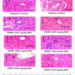 |
Figure 1: Liver histopathology of negative, positive, and carbendazim rats administrated with different concentrations of water kumquat extracts; HWEK: Hot water extracts of kumquat, CWEK: Cold water extracts of kumquat. |
Conclusion
Kumquat water extracts show promise in protecting against carbendazim (CBZ)-induced liver damage. The high phenolic and flavonoid content, especially eriocitrin and hesperidin, contributes to the antioxidant and protective effects. Hot water kumquat extracts were particularly effective in counteracting CBZ-induced liver damage, thanks to their potent antioxidant properties. The study suggests that increased kumquat ingestion may be beneficial for individuals exposed to pesticides, such as farm workers. Kumquat extracts reduced liver damage markers and preserved liver tissue integrity. Higher concentrations of kumquat extracts provided more significant protective benefits. These insights support the potential of kumquat extracts as a natural therapeutic agent against pesticide-induced liver damage and recommend further research and practical application.
Acknowledgement
The authors extend their appreciation to the Researchers supporting project number (RSP2024R349) at King Saud University, Riyadh, Saudi Arabia.
Funding Sources
The author(s) received no financial support for the research, authorship, and/or publication of this article.
Conflict of Interest
The authors do not have any conflict of interest.
Data Availability Statement
This statement does not apply to this article.
Ethics Statement
This study adhered to ethical standards concerning the treatment of animal subjects. The authors obtained ethical approval (number MUFHE/S/NFS/3/22) from the Institutional Animal Care and Use Committee (IACUC) at Menoufia University, Egypt, prior to commencing the experiments. This approval confirms that the study’s protocol was reviewed and sanctioned by an independent ethics committee, ensuring the humane treatment of animals and compliance with established guidelines.
Informed Consent Statement
This study did not involve human participants, and therefore, informed consent was not required.
Permission to Reproduce Material from other Sources
Not applicable
Clinical Trial Registration
This research does not involve any clinical trials.
Author Contributions
- Heba Ezz El-Din Yossef – Conceptualization, Methodology, Writing – Original Draft.
- Mohamed Mohamed Badr – Data Collection, Analysis
- Mohamed Farouk Elsadek – Funding Acquisition, Resources
- Khalid Suliman Al-Numair– Funding Acquisition, Resources
- Abeer Ahmed Khedr– Writing – Review & Editing
- Hend Awad Allah El Sedody – Conceptualization, Methodology, Writing – Original Draft, Data Collection,
- Sunita Singh – Writing – Review and Editing
- Amin Abd El Halim Kandil – Writing – Review & Editing
- Ayman Younes Allam – Conceptualization, Methodology, Writing – Original Draft, Data Collection, Analysis, Writing – Review & Editing, Visualization, Supervision, Project Administration.
References
- Veerappan M, Hwang I, Pandurangan M. Effect of cypermethrin, carbendazim and their combination on male albino rat serum. International Journal of Experimental Pathology. 2012;93(5):361-369.
CrossRef - Wu S, Jin C, Wang Y, Fu Z, Jin Y. Exposure to the fungicide propamocarb causes gut microbiota dysbiosis and metabolic disorder in mice. Environmental Pollution. 2018;237:775-783.
CrossRef - Yang G, Wang Y, Li J, Y Wang, J Li, D Wang, Z Bao, Q Wang, Y Jin. Health risks of chlorothalonil, carbendazim, prochloraz, their binary and ternary mixtures on embryonic and larval zebrafish based on metabolomics analysis. Journal of Hazardous Materials. 2021;404:124240.
CrossRef - Riseh RS, Vatankhah M, Hassanisaadi M, Tamanadar E, Skorik Y. Nanocomposites in agriculture as pesticides for plant protection: a review. Advances in Natural Sciences: Nanoscience and Nanotechnology. 2024;15(2):023003.
CrossRef - Balkrishna A, Kumari A, Kumar A, A. Vedpriya Chauhan, Ankush Upadhyay, Navneet Kumar Guleria, Ishita Amarowicz, Ryszard Kumar, Dinesh Kuca, Kamil. Biosensors for detection of pesticide residue, mycotoxins and heavy metals in fruits and vegetables: A concise review. Microchemical Journal. 02024; 111-292.
- Leskovac A, Petrović S. Pesticide use and degradation strategies: food safety, challenges and perspectives. 2023;12(14):2709.
CrossRef - Fenta L, Mekonnen H. Microbial biofungicides as a substitute for chemical fungicides in the control of phytopathogens: current perspectives and research directions. 2024;2024(1):5322696.
CrossRef - Brauer VS, Rezende CP, Pessoni AM, Rezende,De Paula, Renato Graciano Rangappa, Kanchugarakoppal S Nayaka, Siddaiah Chandra Gupta, Vijai Kumar Almeida, Fausto. Antifungal agents in agriculture: Friends and foes of public health. 2019;9(10):521.
CrossRef - Renuka N, Guldhe A, Prasanna R, Singh P, Bux F. Microalgae as multi-functional options in modern agriculture: current trends, prospects and challenges. Biotechnology advances. 2018;36(4):1255-1273.
CrossRef - Kumar A, Rustagi S, Malik S, Choudhary, Priyvart Khan, Zakir Showkat, Ayman Younes Allam Naik, Bindu Kumar, Vijay Chaudhary, Vishal . Highly potent antioxidant/antibacterial biogenic zno nanoparticles-enabled nano-scavenger reinforced by Aegle Marmelos (Linn) Rind’s extract. ECS Journal of Solid State Science and Technology. 2023;12(7):077003.
CrossRef - Rahman MU, Liu X, Wang X, Fan B. Grapevine gray mold disease: infection, defence and management. Horticulture Research. 2024:uhae182.
CrossRef - Al Jumayi HA, Allam AY, El-Beltagy AE-D, Algarni EH, Mahmoud SF, El Halim Kandil AA. Bioactive compound, antioxidant, and radical scavenging activity of some plant aqueous extracts for enhancing shelf life of cold-stored rabbit meat. 2022;11(6):1056.
CrossRef - Cortés I, di Liberto MG, Kaufman TS, Derita MG, Bracca AB. Synthesis and evaluation of aromatic methoxime derivatives against five postharvest phytopathogenic fungi of fruits. Main structure–activity relationships. Food chemistry. 2020;321:126701.
CrossRef - Karrar E, Ahmed IAM, Wei, Wei Sarpong, Frederick Proestos, Charalampos Amarowicz, Ryszard Oz, Emel Sheikha, Aly Farag., Ayman Y. Allam Oz, Fatih. Characterization of volatile flavor compounds in supercritical fluid separated and identified in gurum (Citrulluslanatus var. colocynthoide) seed oil using HSME and GC–MS. 2022;27(12):3905.
CrossRef - Singh S, Nasib Singh, N. S., Vijay Kumar, V. K., Shivika Datta, S. D., Wani, A. B., Damnita Singh, D. S.,& Joginder Singh, J. S. Toxicity, monitoring and biodegradation of the fungicide carbendazim. Environmental chemistry letters. 2016;14(3):317-329.
CrossRef - Sharma M, Maheshwari N, Khan FH, Mahmood R. Carbendazim toxicity in different cell lines and mammalian tissues. Journal of Biochemical and Molecular Toxicology. 2022;36(12):e23194.
CrossRef - Antonio M, Alcaraz MR, Culzoni MJ. Advances on multiclass pesticide residue determination in citrus fruits and citrus-derived products–A critical review. Environmental Science and Pollution Research. 2024:1-24.
CrossRef - Kumari S, Sundar S, Rustagi S, Allam AY. Technological Advancement in the Development of Functional Food. Functional Foods: CRC Press:26-48.
CrossRef - Abolaji A, Awogbindin I, Adedara I, Farombi E. Insecticide chlorpyrifos and fungicide carbendazim, common food contaminants mixture, induce hepatic, renal, and splenic oxidative damage in female rats. Human & experimental toxicology. 2017;36(5):483-493.
CrossRef - Chen Y, Yang J, Yao B, Zhi D, Luo L, Zhou Y. Endocrine disrupting chemicals in the environment: Environmental sources, biological effects, remediation techniques, and perspective. Environmental Pollution. 2022:119918.
CrossRef - Allam A. Effect of addition loquat (eribotrya japonica) seed powder extract as a natural bioactive compound on the quality characteristics of goat meat nuggets during refrigerated storage–Part I. Journal of Food and Dairy Sciences. 2023;14(3):51-62.
CrossRef - Singh S, Sharma AC, Chaurasia PK, Kumar V, Bharati SL, Allam AYF. Prospects and Importance of Training Needs in Peer Review Models. Scientific Publishing Ecosystem: An Author-Editor-Reviewer Axis: Springer; 2024:347-365.
CrossRef - Zhang , Zhang, S., Wu, R., Zuo, W., Timofte, R., Xing, X., … & Wu, P.. NTIRE 2024 Challenge on Bracketing Image Restoration and Enhancement: Datasets Methods and Results. Paper presented at: Proceedings of the IEEE/CVF Conference on Computer Vision and Pattern Recognition2024.
- Lin H, Zhou L, Lu S, Yang H, Li Y, Yang X. Occurrence and spatiotemporal distribution of natural and synthetic steroid hormones in soil, water, and sediment systems in suburban agricultural area of Guangzhou City, China. Journal of Hazardous Materials. 2024;470:134288.
CrossRef - Pimentão AR, Cuco AP, Pascoal C, Cássio F, Castro BB. Current trends and mismatches on fungicide use and assessment of the ecological effects in freshwater ecosystems. Environmental Pollution. 2024:123678.
CrossRef - Ozcan-Sinir G, Ozkan-Karabacak A, Tamer CE, Copur OU. The effect of hot air, vacuum and microwave drying on drying characteristics, rehydration capacity, color, total phenolic content and antioxidant capacity of Kumquat (Citrus japonica). Food Science and Technology. 2018;39:475-484.
CrossRef - Özkan-Karabacak A, Özcan-Sinir G, Çopur AE, Bayizit M. Effect of osmotic dehydration pretreatment on the drying characteristics and quality properties of semi-dried (Intermediate) Kumquat (Citrus japonica) slices by vacuum dryer. 2022;11(14):2139.
CrossRef - Venkatachalam , Charoenphun, N., Srean, P., Yuvanatemiya, V., Pipatpanukul, C., Pakeechai, K.,& Wongsa, J.. Phytochemicals, Bioactive Properties and Commercial Potential of Calamondin (Citrofortunella microcarpa) Fruits: A Review. Molecules. 2023;28(8):3401.
CrossRef - Kandil A, Aly-Aldin M, Allam A. Quality characteristics of processed low-fat beef sausage as affected by chickpea protein isolates prolonged cold storage. Journal of Food and Dairy Sciences. 2020;11(12):363-368.
CrossRef - Ziogas V, Ganos C, Graikou K, Cheilari A, Chinou I. Chemical Analyses of Volatiles from Kumquat Species Grown in Greece—A Study of Antimicrobial Horticulturae. 2024;10(2):131.
CrossRef - Liu Y, Liu Y, Liu Y, Liu H, Shang Y. Evaluating effects of ellagic acid on the quality of kumquat fruits during storage. Scientia Horticulturae. 2018;227:244-254.
CrossRef - Lou S-N, Lai Y-C, Hsu Y-S, Ho C-T. Phenolic content, antioxidant activity and effective compounds of kumquat extracted by different solvents. Food chemistry. 2016;197:1-6.
CrossRef - Gurjar VK, Shrivastava V, Pal D. Effective Treatment and Control of Anti-Proliferative Diseases with Orange and Kumquat Seeds: Description, Chemistry and Uses. Seeds: Anti-proliferative Storehouse for Bioactive Secondary Metabolites: Springer; 2024:583-603.
CrossRef - Olcay N, Demir MK. Effect of kumquat (Fortunella margarita) powders dried by different methods on some physical and chemical properties of cake. Journal of Food Measurement and Characterization. 2021;15(6):5360-5368.
CrossRef - Abdallah IZ, Ahmed M, Montaser S, Hafez SS. Efficiency of Kumquat fruit (Fortunella margarita) extract against hepatotoxicity and infertility induced by Gamma irradiation in male albino rats. Egyptian Journal of Radiation Sciences and Applications. 2019;32(2):187-199.
CrossRef - Lou S-N, Ho C-T. Phenolic compounds and biological activities of small-size citrus: Kumquat and calamondin. Journal of Food and Drug Analysis. 2017;25(1):162-175.
CrossRef - Olcay N, Demir MK. Effect of kumquat (Fortunella margarita) powders dried by different methods on some physical and chemical properties of cake. Journal of Food Measurement and Characterization. 2021:1-9.
CrossRef - Fuloria, S., Mehta, J., Chandel, A., Sekar, M., Rani, N. N. I. M., Begum, M. Y., … & Fuloria, N. K. . A comprehensive review on the therapeutic potential of Curcuma longa Linn. in relation to its major active constituent curcumin. Frontiers in Pharmacology. 2022;13:820806.
CrossRef - Paiva LS, Motta MH, Baptista JAB. Nutraceutical Value of Eleven Aromatic Medicinal Plants and Azorean Camellia Sinensis: Comparison of Antioxidant Properties and Phenolic and Flavonoid Contents. 2024;12(7):1375.
CrossRef - Berridge MV, Herst PM, Tan AS. Tetrazolium dyes as tools in cell biology: new insights into their cellular reduction. Biotechnology annual review. 2005;11:127-152.
CrossRef - Bakre DS, Kaliwal BB. In-vitro Assessment of Carbendazim and Copper oxychloride cytotoxicity on HaCaT and HepG2 human cell lines. Journal of Applied Biology & Biotechnology Vol. 2017;5(03):023-029.
- FM E-T, OA M, Shadia A, Ghada R. Biological evaluation for some plants used for treatment of diabetes and hyperlipidaemia in rats. Journal of Environmental Science. 2016;35(1):91-111.
CrossRef - Reeves PG, Nielsen FH, Fahey Jr AIN-93 purified diets for laboratory rodents: final report of the American Institute of Nutrition ad hoc writing committee on the reformulation of the AIN-76A rodent diet. The Journal of nutrition. 1993;123(11):1939-1951.
CrossRef - Heiðarsdóttir ÍS. Determination of half maximal inhibitory concentration (IC50) of several anticancer drugs for inhibiting CYP3A4 metabolism of midazolam 2020.
- Reeves PG, Nielsen FH, Fahey Jr GC. AIN-93 purified diets for laboratory rodents: final report of the American Institute of Nutrition ad hoc writing committee on the reformulation of the AIN-76A rodent diet. Oxford University Press; 1993.
CrossRef - Bergmeyer H, Harder M. A colorimetric method of the determination of serum glutamic oxaloacetic and glutamic pyruvic transaminase. Clin Biochem. 1986;24(48):1-488.
- Kachmar J, Moss D. Enzymes. IN Fundamentals of Clinical Chemistry. NW Tietz, Editor. Saunders, Philadelphia; 1976.
- Varley H. Practical clinical biochemistry. Practical clinical biochemistry. 1954.
- Reinhold Total protein, albumin, and globulin. Standard methods of clinical chemistry. 1953;1:88-97.
CrossRef - Burtis C, Ashwood E, Border B. Liver functions. Tietz Fundamentals of Clinical Chemistry, 5th (ed), Saunders Company, Philadelphia. 2001:748-770.
CrossRef - Cashore WJ. Free bilirubin concentrations and bilirubin-binding affinity in term and preterm infants. The Journal of pediatrics. 1980;96(3):521-527.
CrossRef - Moron MS, Depierre JW, Mannervik B. Levels of glutathione, glutathione reductase and glutathione S-transferase activities in rat lung and liver. Biochimica et biophysica acta (BBA)-general subjects. 1979;582(1):67-78.
CrossRef - Nishikimi M, Rao NA, Yagi K. The occurrence of superoxide anion in the reaction of reduced phenazine methosulfate and molecular oxygen. Biochemical and biophysical research communications. 1972;46(2):849-854.
CrossRef - Babson AL, Babson SR. Kinetic colorimetric measurement of serum lactate dehydrogenase activity. Clinical chemistry. 1973;19(7):766-769.
CrossRef - Young I. Measurement of total antioxidant capacity. BMJ Publishing Group; 2001.
CrossRef - Carleton HM, Haynes F. Histological technique. Vol 2: Oxford University Press; 1926.
CrossRef - Carleton HM, Drury RAB, Wallington EA. Carleton’s histological technique. Oxford University Press, USA; 1980.
- Sangeeta A, Gopalakrishnan K, Mishra P. New Food Product Development from Citrus Fruits. Citrus Fruits and Juice: Processing and Quality Profiling: Springer; 2024:223-258.
CrossRef - Zhou X, Zhang X, Liu X, Ji X, Zhang Q, Yang X. Effects of different drying techniques on sea buckthorn pomace: Comprehensive assessment of drying characteristics, physicochemical properties, and odor. Frontiers in Sustainable Food Systems. 2024;8:1434121.
CrossRef - Arnold M, Gramza-Michalowska A. Recent Development on the Chemical Composition and Phenolic Extraction Methods of Apple (Malus domestica)—A Review. Food and Bioprocess Technology. 2024;17(9):2519-2560.
CrossRef - Boateng ID. Mechanisms, capabilities, limitations, and economic stability outlook for extracting phenolics from agro-byproducts using emerging thermal extraction technologies and their combinative effects. Food and Bioprocess Technology. 2024;17(5):1109-1140.
CrossRef - Rajan M, de Carvalho Batista T, Santos de Oliveira C, de Oliveira DG, Narain N. Optimization of solvent extraction and HPLC-DAD method parameters for determination of phenolic compounds in various Brazilian propolis. Journal of Apicultural Research. 2024;63(3):556-569.
CrossRef - Grecco KD, Santos, K. R., Aragão, F. B., Galter, I. N., Lascola, M. B., Dos Santos, S. N., & Matsumoto, S. T.. Toxicogenetic, biochemical, and physiological effects of azoxystrobin and carbendazim fungicides over Lactuca sativa L. and Phaseolus vulgaris L. Environmental Science and Pollution Research. 2024;31(31):44036-44048.
CrossRef - Mabrouk NE, Mastouri M, Lizard G, Aouni M, Harizi H. In vitro immunotoxicity effects of carbendazim were inhibited by n-acetylcysteine in microglial BV-2 cells. Toxicology in Vitro. 2024;97:105812.
CrossRef - Hashim M, Al-Attar AM, Alomar MY, Omar AMS, Alkenani NA, Zeid IMA. Alleviation of carbendazim toxicity effect by Moringa oleifera oil and Linum usitatissimum L. oil on testes of male rats: Physiological, histological and in silico study. Saudi Journal of Biological Sciences. 2024;31(2):103921.
CrossRef - Abdallah N, Ahlan AR, Abdullah OA. The role of quality factors on learning management systems adoption from instructors’ perspectives. The Online Journal of Distance Education and e-Learning. 2019;7(2):133.
- Wang K, Chen X. Protective effect of flavonoids on oxidative stress injury in Alzheimer’s disease. Natural Product Research. 2024:1-28.
CrossRef - Shukla SA, Bachireddy, P., Schilling, B., Galonska, C., Zhan, Q., Bango, C., & Wu, C. J. (. Cancer-germline antigen expression discriminates clinical outcome to CTLA-4 blockade. 2018;173(3):624-633. e628.
CrossRef - Hashem HE, Attia MH, El Wahed Hasan EA, Abdou DM. Valuable Role of Beta-D-Glucan in The Diagnosis of Invasive Fungal Infections in Patients with Hematological Malignancies. Egyptian Journal of Hospital Medicine. 2024;95.
CrossRef - Mahmoud M, Gobet N, Cruz-Dávalos DI, Mounier N, Dessimoz C, Sedlazeck FJ. Structural variant calling: the long and the short of it. Genome biology. 2019;20:1-14.
CrossRef - Madboli AE-NA, Seif MM. Adiantum capillus-veneris Linn protects female reproductive system against carbendazim toxicity in rats: immunohistochemical, histopathological, and pathophysiological studies. Environmental Science and Pollution Research. 2021;28(16):19768-19782.
CrossRef - Mahmoud AM, Hernandez Bautista RJ, Sandhu MA, Hussein OE. Beneficial effects of citrus flavonoids on cardiovascular and metabolic health. Oxidative medicine and cellular longevity. 2019;2019.
CrossRef - Kashyap A, Cook K, Behera MD. Geomorphic imprint of high mountain floods: Insight from the 2022 hydrological extreme across the Upper Indus terrain in NW Himalayas. 2024;2024:1-36.
CrossRef

This work is licensed under a Creative Commons Attribution 4.0 International License.


