
Introduction
Kaempferia angustifolia, commonly known as temu kunci or white turmeric, is a medicinal plant in the Zingiberaceae family, which comprises approximately 60 species of small rhizomatous herbs.1 Native to East Tropical Asia, this genus is characterized by small rhizomatous herbs with short rhizomes and thick, aromatic tuberous roots.2 While Kaempferia angustifolia is less prominent in Malaysia compared to its relatives K. galanga and K. rotunda3, it is recognized in Indonesia by names such as Kunci pepet, Kunci menir, and Kunci kunot, and in Vietnam as Thao Nhang Haeng.1
Traditionally, the rhizomes of K. angustifolia are prized for their pleasant aroma and are widely used in folk medicine to treat ailments such as colds, stomachaches, coughs, diarrhea, fever, and dysentery.4 Recent studies have revealed the therapeutic potential of edible Southeast Asian plants, including K. angustifolia, for applications such as cancer chemoprevention and antimicrobial activity.3 Additionally, K. angustifolia has demonstrated biological activities, including antioxidant, antimicrobial, anti-obesity, and anti-allergic effects.4 Despite these promising properties, research on K. angustifolia as an anti-obesity agent remains limited. In Indonesia, the rhizomes are traditionally used in slimming remedies, prompting further investigation into their potential to regulate adipocyte differentiation in murine 3T3-L1 cells.4,5
The essential oil of K. angustifolia has shown moderate antimicrobial activity, particularly against Pseudomonas aeruginosa (10.63 ± 0.23 mm and 10.03 ± 0.22 mm inhibition zones), but it exhibits minimal efficacy against pathogens such as Escherichia coli, Bacillus subtilis, Streptococcus faecalis, Candida albicans, and Microsporum gypseum.6 Extracts and compounds from Kaempferia species have demonstrated diverse therapeutic applications, including inhibition of Staphylococcus aureus and Pseudomonas spp., treatment of knee osteoarthritis, and inhibition of breast cancer resistance protein (BCRP). They also exhibit anti-inflammatory, anti-acne, and anticholinesterase activities, support wound healing, mitigate obesity-induced dermatopathy, and combat drug-resistant strains of Mycobacterium tuberculosis. Additionally, Kaempferia species possess neuroprotective and anti-nociceptive properties and have shown activity against HIV-1, in vitro anti-allergenic effects, and larvicidal activity against Aedes aegypti.7
Specific compounds from K. angustifolia, such as 20-hydroxy-4,4,6-trimethoxychalcone and 25-dien-3-ol, exhibit cytotoxic effects against human cancer cell lines, including HL-60 (promyelocytic leukemia) and MCF-7 (breast cancer).4 The Zingiberaceae family, known for its antimicrobial properties, produces extracts and essential oils effective against bacteria, fungi, and yeasts. Antioxidant compounds in K. angustifolia include boesenboxide, crotepoxide, 2′-hydroxy-4,4′,6′-trimethoxychalcone, kaempfolienol, and zeylenol, alongside catechin, kaempferol, quercetin, gallic acid, caffeic acid, and α-tocopherol, which are particularly notable for their anticancer and antioxidant properties. This species is a rich source of secondary metabolites, including diterpenoids, steroids, saponins, cyclohexane oxides, flavonoids, and chalcones.8
These metabolites have applications in medicine, nutrition, beverages, fragrances, cosmetics, and other industries.9 Chalcones, characterized by small molecular structures, act as Michael acceptors, enabling covalent interactions with proteins that drive biological activity.10 The composition and concentration of these metabolites vary with factors such as growth environment, harvest season, plant part used, and solvent properties, directly influencing their biological activity.11 Flavonoids in K. angustifolia include glycosidic and aglycone types. C-glycosidic flavonoids, with sugar substituents at C(6) and/or C(8) of the aglycone, are of particular interest. Their moderate polarity and thermolability make reversed-phase liquid chromatography (LC) a preferred analysis method, with advanced techniques like LC-MS and LC-MS/MS providing precise identification of flavonoids and their substituent positions.9
In silico pharmacology, a growing field leveraging computational models to analyze biological and medical data, facilitates the prediction and discovery of therapeutic compound.12 Previous studies on K. angustifolia have analyzed its chemical composition and biological activities, with metabolomics revealing changes in metabolite profiles under various conditions.13 Techniques like LC-MS/MS provide the sensitivity and selectivity needed for detailed metabolite profiling.
In overall, the research on Kaempferia angustifolia and FemiVine V Jelly highlights their antioxidant and cytotoxic potential, yet certain gaps remain. While the study demonstrates the superior antioxidant activity of FemiVine V Jelly and the stronger cytotoxic effect of the raw extract on MDA-MB-231 breast cancer cells, further investigation is needed to understand the mechanisms underlying these differences. Additionally, the in-silico analysis suggests promising pharmacokinetic properties for vitexin and β-sitosterol, but their selective cytotoxicity against normal fibroblasts remains unexplored. Future studies should focus on optimizing flavonoid extraction, evaluating potential synergistic effects, and conducting in vivo experiments to validate their therapeutic efficacy. Thus, this study aims to evaluate the antioxidant properties, cytotoxic potential, and pharmacokinetic characteristics of flavonoids in K. angustifolia extracts, focusing on their therapeutic potential against MDA-MB-231 breast cancer cells. The innovative FemiVine V jelly product and raw extracts are analyzed as part of this investigation.
Materials and Methods
Materials and Chemicals
The following materials were used in this study: Folin-Ciocalteu reagent, sodium nitrite, catechin, gallic acid, Sephadex G-25, and MTT (3-[4,5-dimethylthiazol-2-yl]-2,5-diphenyltetrazolium bromide), acetate buffer (pH 3.6),10 mM 2,4,6-tris(2-pyridyl)-s-triazine (TPTZ) , hydrochloric acid (HCl), iron(III) chloride hexahydrate (FeCl₃·6H₂O) all obtained from Sigma Chemical Co. (St. Louis, MO, USA). LH-20 was procured from Amersham Biosciences (Uppsala, Sweden), and Whatman No. 1 filter paper was purchased from Fisher Scientific (Fair Lawn, NY, USA). High-performance liquid chromatography, HPLC-grade acetonitrile and acetic acid were supplied by Merck (Darmstadt, Germany). Two types of samples were analyzed: the raw extract and the FemiVine V jelly commercial product. The raw extract was in powder form, the process of drying K. angustifolia were washed and cut into small slices then put onto the aluminum tray and oven-dried at 50 ∘C for 24 hrs. The dried samples were then ground to powder form and kept in bottles prior to extraction process. The raw extract and FemiVine V jelly were similarly dissolved at the same concentration. Both samples were centrifuged at 10,000 rpm for 10 minutes, and the resulting supernatant was filtered using a 0.22 µm syringe filter. The filtered solutions were then transferred into 20 µL inserts prior to injection into the Liquid Chromatography/Mass Spectrometry (LC/MS) system.
Phytochemicals Molecular Profiling Analysis
The Liquid Chromatography-Quadrupole Time-of-Flight Mass Spectrometry (LC/MS-QTOF) system employed in this study comprised an Agilent 1200 liquid chromatography system equipped with a binary pump, a vacuum degasser unit, an auto-sampler, and a 6520-quadrupole time-of-flight mass spectrometer with an electrospray ionization (ESI) source. Chromatographic separation was carried out using an Agilent ZORBAX Eclipse Plus C18 Rapid Resolution HT column (2.1 × 100 mm, 1.8 µm particle size) maintained at a temperature of 40 °C. The mobile phase consisted of two components: (A) 0.1% formic acid in deionized water and (B) 0.1% formic acid in acetonitrile. The gradient elution program was as follows: a) 0.00–18.00 minutes: Linear gradient from 5% (B) to 95% (B); 18.00–23.00 minutes: Held at 95% (B) and c) 23.01 minutes: Returned to 5% (B). The total run time was 30 minutes, with the system pre-equilibrated for 2 minutes before each new injection. A sample injection volume of 2 µL was used, and the mobile phase flow rate was maintained at 0.25 mL/min. The mass spectrometer operated in positive ESI mode under the following conditions: a) Gas temperature: 325 °C; b) Gas flow rate: 11 L/min; c) Nebulizer pressure: 35 psi. Data were analyzed using Agilent Mass Hunter Qualitative Analysis software (version B.05.00, Agilent Technologies, Santa Clara, CA, USA). Chromatographic profiles were assessed based on the accurate mass measurements obtained from the LC/MS system.14, 15
Determination of Total Phenolic Contents
The total phenolic content of the raw Kaempferia angustifolia extract and FemiVine V jelly was quantified using a modified Folin-Ciocalteu method. A 125 µL aliquot of a known dilution of the extract was transferred into a test tube and mixed with 0.5 mL of Folin-Ciocalteu reagent. The mixture was vortexed for 15 seconds and left to stand at room temperature (20 °C) for 6 minutes. Subsequently, 1.25 mL of a 7% sodium carbonate solution was added to the mixture, and the volume was adjusted to 3 mL using distilled, deionized water. The reaction mixture was allowed to develop color over 90 minutes. Absorbance was then measured at 760 nm using a Hewlett-Packard UV-Vis spectrophotometer (Palo Alto, CA, USA). The total phenolic content was calculated by comparing the absorbance values to a standard curve prepared with gallic acid solutions, and the results were expressed as milligrams of gallic acid equivalents (mg GAE). All measurements were conducted in triplicate, and the results are presented as the mean of three determinations (n = 3).16
DPPH Radical Scavenging
A 0.1 mL aliquot of the acetone extract prepared earlier was combined with 3.9 mL of an 80% ethanolic 0.6 mM DPPH solution in a test tube. The mixture was vortexed for 15 seconds and allowed to stand for 180 minutes at room temperature.17 After this incubation period, the absorbance was measured at 515 nm using a Hewlett-Packard UV/Vis spectrophotometer (Palo Alto, CA, USA). Under these conditions, most tested compounds are expected to react completely within 180 minutes, while vitamin C, due to its rapid oxidation, reacts in less than 1 minute. An 80% ethanol solution was used as the blank, while the control consisted of the DPPH solution without any test sample (3.9 mL of DPPH mixed with 0.1 mL of 80% ethanol). All tests were performed in triplicate (n = 3). The antioxidant activity of the samples was evaluated using two parameters: a) Median Effective Concentration (EC50): The concentration of antioxidant (test sample) required to achieve a 50% reduction in the absorbance of DPPH radicals, expressed as total phenolics (mg); b) Scavenging Activity (%): Calculated using the formula:
Inhibition (%) of DPPH absorbance = (Acontrol – Atest)/Acontrol x 100 —————– (1)
A dose-response curve (absorbance of DPPH vs. concentration of antioxidants) was plotted to establish standard curves and calculate the EC50. Vitamin C was used as the standard reference, and the results were expressed as vitamin C equivalent antioxidant capacity (VCEAC).
FRAP Assay
The reducing capacity of the sample was evaluated using the FRAP (Ferric Reducing Antioxidant Power) method, as described by Elshamy et al. (2019).7 Briefly, the FRAP reagent was freshly prepared by mixing the following components: a) 300 mM acetate buffer (pH 3.6), b) 10 mM 2,4,6-tris(2-pyridyl)-s-triazine (TPTZ) dissolved in 40 mM hydrochloric acid (HCl), and c) 20 mM iron(III) chloride hexahydrate (FeCl₃·6H₂O). A 1.0 mL aliquot of the FRAP reagent was combined with 50 µL of the sample or Trolox standard. The mixture was incubated in the dark at room temperature for 30 minutes to allow the reaction to develop. The reducing capacity of the sample was then determined by measuring absorbance at 595 nm using a spectrophotometer (BMG Labtech).17
2-D Cell Culture and MTT Viability Assay
The MDA-MB-231 adenocarcinoma cell line was cultured in Dulbecco’s Modified Eagle Medium (DMEM), as described by Kim et al. (2014).18 The medium was supplemented with 10% heat-inactivated fetal bovine serum (FBS) and 1% penicillin-streptomycin (P/S). The cells were maintained in a humidified incubator at 37°C with 5% CO₂. Adherent BV-2 cells, at ≥70% confluency, were detached from the flask by trypsinization and subcultured every 72 hours. For the 3-(4,5-dimethylthiazol-2-yl)-2,5-diphenyl-2H-tetrazolium bromide (MTT) assay, 180 µL of BV-2 cells at a density of 20,000 cells per well were seeded into a 96-well plate and incubated for 24 hours. Subsequently, the cells were treated with 20 µL of the following: a) Medium alone, b) 0.01% dimethyl sulfoxide (DMSO), c) Donepezil, or d) labelled dextran (FD) at various concentrations (0.1 µg/mL, 1 µg/mL, 10 µg/mL, and 100 µg/mL). After a further 24-hour incubation period, 50 µL of MTT reagent was added to each well, followed by incubation at 37 °C for 4 hours. Following this, the supernatants were carefully removed, and 100 µL of DMSO was added to each well to dissolve the formazan crystals.19 Absorbance was measured at 550 nm using a microplate reader (Molecular Devices, USA). The normalized viability (%) of the cells was calculated using SoftMax® Pro Software for accuracy and reliable data acquisition and analysis.
In Silico Metabolism, and Absorption, Distribution Excretion (ADME) Prediction and Drug-Likeness Assessment
The QikProp 4.4 software was employed for absorption, distribution, metabolism, and excretion (ADME) predictions. Recognized for its accuracy, simplicity, and efficiency, this program provides key physical descriptors and relevant pharmaceutical properties of active molecules (QikProp 4.4 User Manual, 2015). The “drug-likeness” of the compounds was evaluated based on Lipinski’s “Rule of Five.” Critical physical properties, including molecular weight, hydrophobicity (logP), the number of hydrogen bond acceptors, and the number of hydrogen bond donors, were analyzed to determine their compliance with the rule. Furthermore, the potential of each compound to cross the blood-brain barrier (BBB) was assessed, as described by Zolkiffly et al. (2021).12
Statistical Analysis
Results are given as mean values ± standard deviation from measurements of three replicates (n = 3). Statistical analysis data can be obtained from Minitab statistical software (Version 22.0, Minitab Inc., United States). The independent t-test was then performed to compare the mean values obtained at a significant level of 95% (p<0.05).
Results
In the present study, four flavonoid compounds were identified in K. angustifolia extracts through Q-TOF LC-MS analysis (Table 1, Figures 1 and 2). These compounds were detected in two samples: FemiVine V jelly and the raw (powdered) species. Identification was achieved using full mass spectra and MS² fragmentation patterns, which were compared with published data and MS² references, such as those from Mass Bank. The base peak chromatograms obtained in positive ionization mode (Figures 1 and 2) revealed distinct metabolite profiles for the two extracts.
The fragmentation patterns of the identified compounds were consistent across samples, especially at low collision energies (<70% eV). For instance, isoorientin (luteolin 6-C-glucose) exhibited the loss of a water molecule, resulting in a fragment at m/z 448 [M–H–18]−. This was observed at retention times of 20.643 minutes in FemiVine V jelly and 23.132 minutes in the raw extract. In contrast, orientin (luteolin 8-C-glucose) did not show this loss of water.
 |
Figure 1: LC-MS Chromatogram of FemiVine V Jelly commercialized product with the number of molecular features (M/Z) detected as of 208 |
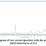 |
Figure 2: LC-MS chromatogram of raw extract (powder) with the number of molecular features (M/Z) detected as of 121 |
Table 1: Selected metabolites profiling of K. Angustifolia extracts FemiVine V jelly and raw extract as identified by LC-MS
|
FemiVine V jelly |
Compound | m/z | Formula | RT (min) | Height |
| Orientin | 447.69 | C21H20O11 | 20.643 |
1119 |
|
|
Vitexin |
432.24 | C21H20O10 | 20.664 | 2048 | |
| Kaempfolienol | 354.5 | C20H34O5 | 1.064 |
3126 |
|
|
β- Sitosterol |
414.7 | C29H50O | 15.793 | 3064 | |
| Raw extract (powder) | Orientin | 447.40 | C21H20O11 | 23.132 |
1004 |
|
Vitexin |
435.36 | C21H20O10 | 20.619 | 2322 | |
| Kaempfolienol | 289.05 | C20H34O5 | 1.062 |
1503 |
|
|
β- Sitosterol |
413.32 | C29H50O | 22.113 |
4695 |
The antioxidant properties of FemiVine V jelly and its corresponding raw extract were assessed using three methods in Table 2: total polyphenol content (expressed as gallic acid equivalents, GAE), the DPPH assay, and the FRAP assay. This study represents the first documented evaluation of the antioxidant potential of extracts and phytochemicals derived from Kaempferia angustifolia, as summarized in Table 2.
Table 2: Antioxidant activity for FemiVine V jelly and species raw of K. angustifolia
|
Antioxidant Activity |
FemiVine V Jelly | Raw Extract (Powder) |
| Total Polyphenol (GAE) mg/g | 31.93 ± 0.20a |
12.00 ± 0.23b |
|
DPPH (% Inhibition) |
67.60 ± 0.15c | 30.54 ± 0.10d |
| FRAP | 5.08 ± 0.01 (mg/100 g ascorbic acid)e |
4.81 ± 0.05 (mg/100 ml VCEAC)f |
a-fMean with different alphabet shows significant difference between samples (p<0.05)
The cytotoxic screening results (see Table 3) demonstrate the antagonistic effect of the isolated compounds from K. angustifolia against the MDA-MB-231 breast cancer cell line. The tested products were found to be inactive, showing IC50 values of 125 mg/mL for FemiVine V jelly and 82 mg/mL for the raw extract. The raw extract exhibited a stronger IC50 value compared to FemiVine V jelly (p<0.05).
Table 3: IC50 values for both commercialized product and raw extract (powder) of K. angustifolia against MDA-MB-231
|
Product |
Nilai IC50 mg/ml |
|
FemiVine V jelly |
125 ± 0.20a |
| Raw extract (powder) |
82 ± 0.06b |
a-bMean with different alphabet shows significant difference between samples (p<0.05)
Furthermore, Figure 3 presents the cell morphology of MDA-MB-231 cells at both low and high densities from ATCC. Figures 4(a) and (b) display cell morphology for negative and positive controls, along with initial post-treatment observations following the administration of FemiVine V jelly at concentrations of 500 mg/mL (c), 250 mg/mL (d), and 125 mg/mL (e). Figures 4(f), (g), and (h) show the morphology for the raw extract. Furthermore, as the concentration of the product increases, cell morphology begins to display signs of interaction, with noticeable stress appearing across the cell colonies. At a concentration of 500 mg/mL, cells exhibit shrinkage and asymmetric membrane development (Figures 4(c) and (f)) for both FemiVine V jelly and the raw extract.
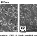 |
Figure 3: Cell morphology of MDA-MB-231 under low and high density from ATCC |
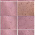 |
Figure 4: Cell morphology for (a) negative control (b) positive control (c) concentration of 500 mg/ml FemiVine V jelly (d) concentration of 250 mg/ml FemiVine V jelly (e) concentration of 125 mg/ml FemiVine V jelly |
The evaluation of drug-likeness results revealed that both vitexin and β-sitosterol displayed ‘drug-like’ properties, satisfying all four criteria of the Rule of Five. In comparison to orientin and kaempferol, vitexin and β-sitosterol demonstrated acceptable blood-brain barrier (BBB) permeability, as illustrated in Figure 5. Table 4 presents the SwissADME web-based predictions for the physicochemical properties, including lipophilicity, pharmacokinetics, drug-likeness, and medicinal chemistry compatibility of these compounds. The in-silico predictions considered various factors, as shown in Figure 5, such as lipophilicity, molecular weight, and water solubility, all falling within acceptable ranges: lipophilicity (-0.7 to +5.0), molecular weight (150 to 500 g/mol), and water solubility (≤6). In contrast, orientin, vitexin, and kaempferol exhibit polar surface areas exceeding the recommended threshold (>130 Ų), which is associated with lower gastrointestinal absorption rates. Additionally, all compounds were predicted to have carbon saturation in Csp3 values within acceptable limits (≥0.25). The number of rotatable bonds, which reflect compound flexibility, also remained within the recommended range (≤9) for these four flavonoid compounds. Furthermore, the results from the boiled egg prediction analysis (Figure 6) evaluated the passive gastrointestinal absorption (HIA) and brain penetration (BBB) of the compounds using WLOGP (lipophilicity) and TPSA (topological polar surface area).
Table 4: In silico analysis for orientin, vitexin, kaempfolienol and β- Sitosterol
|
Parameter |
Orientin | Vitexin | Kaempfolienol | β-Sitosterol |
| Lipophilicity (XLOGP3) | -0.60 | 1.62 | -5.89 |
8.79 |
|
Size (MV) |
434.35 g/mol | 432.38 g/mol | 364.35 g/mol | 400.68 g/mol |
| Polarity (TPSA) | 201.28 | 181.05 | 202.30 |
20.23 |
|
Solubility (Log S) |
-3.16 | -3.57 | 2.31 | -9.10 |
| Water soluble | Soluble | Soluble | High soluble |
Poorly soluble |
|
Saturation Csp3 |
0.25 | 0.29 | 0.87 | 0.93 |
| Num.rotatable bonds | 2 | 3 | 1 |
6 |
|
GI absorption |
Low | Low | Low | Low |
| BBB permeant | No | No | No |
No |
|
P-gp substrate |
No | No | Yes | No |
| Lipinski (Drug likeness) | No | Yes | Yes |
Yes |
|
Ghose (Drug likeness) |
No | Yes | No | No |
| Bioavailability Score | 0.17 | 0.55 | 0.55 |
0.55 |
|
Synthetic accessibility |
5.12 | 5.19 | 5.3 |
6.17 |
*MW: molecular weight; GI: gastrointestinal; BBB: blood-to-brain barrier; P-gp: Glycoprotein Substrate XLOP3 Lipophilicity between -0.7 and +5.0 Size: MW between 150 and 500 g/mol; Solubility: TPSA between 20 and 130 Å2; solubility: Log S ≤ 6; Saturation: fraction of carbon in sp3 hybridization ≥ 0.25; Flexibility: ≤ 9 rotatable bonds Synthetic Accessibility: 1-10 (very easy to very difficult to synthesize)
log S not higher than 6, saturation : carbon fraction in sp3 hybridization not less than 0.25, and flexibility: not more than 9 rotatable bonds this example, the compound is predicted to be unavailable orally, because it is too flexible and too polar.
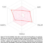 |
Figure 5: The bioavailability radar allows a first look at the drug-likeness of a molecule. The pink area represents the optimal range for each property (lipophilicity: XLOGP3 between -0.7 and +5.0, size: |
 |
Figure 6: Prediction analysis of boiled eggs to estimate the gastrointestinal absorption and brain penetration of vitexin and β- Sitosterol molecules. *Blood-brain barrier (BBB) penetration, |
Discussion
Flavonoid Identification and Glycosylation Patterns in K. angustifolia
Flavonoids are synthesized through a series of condensation reactions involving hydroxycinnamic acid, which interacts with the B-ring and carbon atoms 2, 3, and 4 of the C-ring, and malonyl residues, which contribute to the A-ring. This process results in a base structure characterized by a C6–C3–C6 backbone. The three-carbon bridge connecting the phenyl rings typically undergoes cyclization, forming a third ring (C-ring). In plants, flavonoids exist in various modified forms, often involving hydroxylation, methylation, and glycosylation. Additional modifications, such as attachment of aromatic or aliphatic acids and groups like sulfate, prenyl, methylenedioxyl, or isoprenyl, can further diversify the flavonoid nucleus and its glycosides.20
Glycosylation plays a crucial role in modifying flavonoids, reducing their reactivity and increasing their water solubility. This modification serves as a protective mechanism in plants, preventing cytoplasmic damage and enabling the safe storage of flavonoids in vacuoles. Glycosylation occurs through the attachment of sugar molecules to the flavonoid core, often via acid-resistant C–C bonds, resulting in the formation of flavonoid C-glycosides. These C-glycosides are categorized into mono-C-glycosylflavonoids, di-C-glycosylflavonoids, and C-glycosylflavonoid-O-glycosides. For flavones and flavonols, glycosylation typically occurs at the 7-hydroxyl group, while C-glycosylation usually takes place at the 6C or 8C positions.
For vitexin, retention times of 20.664 minutes in FemiVine V jelly and 20.619 minutes in the raw species were recorded. The molecular ion [M + H]+ was detected at m/z 432.24 and 435.36, with a molecular formula of C21H20O10. Fragmentation primarily involved the sequential loss of C4H8O3, water, and carbon monoxide, resulting in fragments [M – C4H8O3 – H], [M – C4H8O3 – H2O – H], and [M – C4H8O3 – CO – H], respectively.21 For β-sitosterol, the molecular ion [2M + H]+ was detected at m/z 414.7 in FemiVine V jelly and 413.32 in the raw extract, corresponding to a molecular formula of C29H5O10.
The compound formed an ammonium adduct, and its fragmentation yielded cholestanol and sitostanol ions, with water loss producing [M + H – H2O]+.22 Retention times were 15.793 minutes for FemiVine V jelly and 22.113 minutes for the raw extract. Kaempferol was identified in the mass chromatograms with m/z values of 354.5 and 289.05, at retention times of 1.064 minutes and 1.062 minutes, respectively. The fragmentation pattern suggested the presence of kaempferol glucoside, with the loss of [M – H]− to [A – H].21
Antioxidant Properties of K. angustifolia Extracts
The total phenolic content in the fresh, edible portion of K. angustifolia was quantified as milligrams of gallic acid equivalents (GAE) per 100 grams. The results revealed that FemiVine V Jelly contained significantly higher levels of phenolic compounds (31.93 ± 0.20 mg/g) compared to the raw extract (12.00 ± 0.23 mg/g, p<0.05). This difference suggests that the processing involved in the production of FemiVine V Jelly may enhance the concentration of bioactive phenolic compounds, which are known for their antioxidant properties.
To further assess the antioxidant capacity, the DPPH (2,2-diphenyl-1-picrylhydrazyl) assay was utilized. This assay evaluates the ability of antioxidants to scavenge free radicals through either a single electron transfer (SET) or a hydrogen atom transfer (HAT) mechanism.23,24 The DPPH radical, a stable free radical, is used as a chromogenic reagent that changes color upon reacting with antioxidants, allowing for the quantification of their scavenging ability. Both the chloroform and methanol extracts of K. angustifolia showed similar effectiveness in scavenging DPPH radicals. However, FemiVine V Jelly exhibited a significantly higher inhibition of 67.6 ± 0.15%, compared to just 30.54 ± 0.15% for the raw extract (p<0.05). This indicates that FemiVine V Jelly has superior radical-scavenging ability, potentially due to its higher concentration of phenolic compounds, which contribute to its enhanced antioxidant activity.
The Ferric Reducing Antioxidant Power (FRAP) assay was also employed to evaluate the antioxidant capacity by measuring the reduction of Fe³⁺-TPTZ (ferric-tripyridyltriazine) to Fe²⁺-TPTZ. This method provides a direct assessment of an antioxidant’s reducing power. FemiVine V Jelly demonstrated stronger reducing power than the raw extract, with values of 5.08 ± 0.01 mg/100 g ascorbic acid compared to 4.81 ± 0.05 mg/100 mL Vitamin C equivalent antioxidant capacity (VCEAC), respectively (p<0.05). This result further supports the observation that FemiVine V Jelly has superior antioxidant activity, making it a potentially valuable source of natural antioxidants.
Taken together, these findings highlight that FemiVine V Jelly not only contains higher levels of phenolic compounds but also exhibits greater antioxidant efficacy than the raw extract. This enhanced antioxidant potential could have implications for the product’s therapeutic use, particularly in mitigating oxidative stress-related diseases, where antioxidant activity plays a critical role. The difference in antioxidant capacity between the jelly and the raw extract underscores the importance of processing methods in improving the bioavailability and effectiveness of natural products. Further exploration of these properties could pave the way for developing more potent antioxidant-rich formulations for health benefits.
Cytotoxicity and Antiproliferative Effects
A recent study examined the antiproliferative effects of these compounds across various cancer cell lines, indicating that structural features such as methoxy substituents, α-methylation of the enone moiety, and 2′-oxygenated substituents contribute to cytotoxic activity due to their antimitotic properties. Additionally, methoxylated chalcones have demonstrated cytotoxic activity by inhibiting tubulin polymerization in multiple tumor cell lines, with IC50 values generally in the high micromolar range (IC50: >50 µM). Phenolic compounds, including flavonoids, are known for their antioxidant properties, as they donate electrons to free radicals, thereby converting them into stable metabolites and halting radical chain reactions.25
This mechanism may contribute to further damage and the eventual death of cancer cells. The observed antiproliferative activity was not solely due to individual compounds but rather a combination of several flavonoids, including vitexin, orientin, kaempferol, and β-sitosterol, all of which are present in the product.26 Although other organic solvents, such as methanolic leaf extracts, have been used to extract these phenolic constituents and have demonstrated significant efficacy in killing cancer cells, subacute oral administration in rats showed no recorded toxicity.19, 23, 27
In Silico Evaluation of Drug-Likeness, BBB Permeability and Therapeutic Potential of Flavonoid Compounds
Several descriptor properties were employed to assess the molecular characteristics of orientin, vitexin, kaempferol, and β-sitosterol, focusing on the ‘Rule of Five’ (ROF) and blood-brain barrier (BBB) permeability.26, 28 The prediction analysis of boiled eggs (Figure 6) indicates that both vitexin and β-sitosterol are likely to be absorbed passively through the gastrointestinal tract but do not appear to penetrate the brain. Additionally, these two flavonoid compounds are not substrates for P-glycoprotein (P-gp), suggesting that they are not actively transported back into the gut after absorption.29,30 In silico analysis thus suggests that vitexin and β-sitosterol possess favourable pharmacokinetic properties for targeting MDA-MB-231 breast cancer cells. Their good absorption profiles, lack of BBB penetration, and non-substrate status for P-gp highlight their potential as promising candidates for therapeutic development.31,32 However, further research is needed to assess their cytotoxic effects on normal fibroblast cells to confirm their clinical suitability. With additional studies, these compounds could emerge as targeted agents with minimal side effects.
Conclusion
In conclusion, the study investigated four key flavonoids, orientin, vitexin, kaempferol, and β-sitosterol found in FemiVine V Jelly and its raw extract, using Q-TOF LC-MS analysis to identify their presence. Using Q-TOF LC-MS analysis, these compounds were successfully identified in both FemiVine V Jelly and the raw extract, with m/z values of 447.69 (orientin), 432.24 (vitexin), 354.5 (kaempferol), and 414.7 (β-sitosterol). Antioxidant potential was evaluated through Total Phenolic Content (TPC), DPPH, and FRAP assays. FemiVine V Jelly exhibited a significantly higher phenolic content (31.93 ± 0.20 mg/g) compared to the raw extract (12.00 ± 0.23 mg/g, p<0.05). In the DPPH radical scavenging assay, FemiVine V Jelly demonstrated superior scavenging activity (67.60 ± 0.15%) compared to the raw material (30.54 ± 0.10%, p<0.05). Similarly, in the FRAP assay, FemiVine V Jelly displayed stronger reducing power (5.08 ± 0.01 mg/100 g ascorbic acid) than the raw extract (4.81 ± 0.05 mg/100 ml Vitamin C, p<0.05). Cytotoxicity screening revealed that compounds isolated from K. angustifolia exhibited antagonistic effects against MDA-MB-231 breast cancer cells. However, the tested products showed limited activity, with IC50 values of 125 ± 0.20 mg/ml for FemiVine V Jelly and 82 ± 0.06 mg/ml for the raw extract. The raw extract demonstrated stronger cytotoxic potential (p<0.05). Additionally, in-silico analysis suggested that vitexin and β-sitosterol possess favorable pharmacokinetic properties, including good gastrointestinal absorption and no brain penetration, making them promising candidates for peripheral cancer treatments such as MDA-MB-231 breast cancer. Their non-substrate status for P-glycoprotein (P-gp) could enhance intracellular retention. However, further studies are required to evaluate their selective cytotoxicity against cancer cells versus normal fibroblast cells. To emphasize practical implications, future studies could explore the development of FemiVine V Jelly or similar formulations as adjuncts to cancer therapies, particularly in peripheral cancers, by assessing their ability to selectively target and kill cancer cells without harming healthy cells. This would be important in optimizing the use of these compounds in clinical settings and advancing their therapeutic application.
Acknowledgement
We are grateful to Universiti Kebangsaan Malaysia (UKM) for the financial support and the Department of Food Sciences, Faculty of Science and Technology, UKM Bangi for allowing this study to be carried out at the Functional Food and Nutritional laboratory. Figure 3 has been reproduced/adapted with permission from https://www.atcc.org/products/htb-26.
Funding Sources
We sincerely thank Universiti Kebangsaan Malaysia (UKM) for providing funding through grant ST-2023-043, which made this study possible.
Conflict of Interest
The author(s) do not have any conflict of interest.
Ethics Statement
This research did not involve human participants, animal subjects, or any material that requires ethical approval.
Informed Consent Statement
This study did not involve human participants, and therefore, informed consent was not required.
Clinical Trial Registration
This research does not involve any clinical trials.
Data Availability Statement
No data were used elsewhere to support this study and it was entirely a new set of data.
Permission to reproduce materials from sources
Not Applicable
Author Contributions
- Farah Nadiah Mohd Fazil: Conceptualization, Methodology, Writing – Original Draft.
- Ikhwan Zakaria: Data Collection, Analysis, Writing – Review & Editing.
- Roslee Rajikan: Funding Acquisition, Supervision, Project Administration.
- Saiful Irwan Zubairi: Writing, Conceptualization, Methodology, Supervision.
- Zalifah Mohd Kasim: Analysis, Writing – Review & Editing.
- Isa Naina Mohamed: Data Collection, Analysis, Writing – Review & Editing.
- Mohd Faizal Sa’aidin: Data Collection, Analysis, Writing – Review & Editing.
References
- Yeap, Y. S. Y., Kassim, N. K., Ng, R. C., Ee, G. C. L., Saiful Yazan, L., Musa, K. H., Antioxidant properties of ginger (Kaempferia angustifolia) and its chemical markers. Int. J. Food Prop. 2017;20(1),1158–1172.
CrossRef - Tran-Trung, H., Thuy, P. T., Thuan, V. T., Ha, N. X., Van Hue, N., Nguyen-Ngoc, H., Nguyen, T. H. D., Tuan, N. H., Van Chen, T., Hien, N. T. T., Giang, L. D., Chemical composition and antimicrobial activity of essential oil obtained from the rhizomes of Kaempferia champasakensis: in vitro and molecular docking studies. Essent. Oil-Bear. Plants. 2023;26(4), 958–969.
CrossRef - Tang, S. W., Sukari, M. A., Neoh, B. K., Yeap, Y. S. Y., Abdul, A. B., Kifli, N., Cheng Lian Ee, G., Phytochemicals from kaempferia angustifolia and their cytotoxic and antimicrobial activities. Biomed Res Int. 2014, 2014:417674. DOI:10.1155/2014/417674.
CrossRef - Rafi, M., Karomah, A. H., Septaningsih, D. A., Trivadila, Rahminiwati, M., Prama Putri, S., Iswantini, D., LC-MS/MS based metabolite profiling and lipase enzyme inhibitory activity of Kaempferia angustifolia with different extracting solvents. Arab. J. Chem. 2022;15(11).
CrossRef - Wichayapreechar, P., Charoenjittichai, R., Prasansuklab, A., Vinardell, M. P., Rungseevijitprapa, W., Exploring the In Vitro Antioxidant, Anti-Aging, and Cytotoxic Properties of Kaempferia galanga Rhizome Extracts for Cosmeceutical Formulations. Cosmetics. 2024;11(3).
CrossRef - Yenjai, C., Prasanphen, K., Daodee, S., Wongpanich, V., Kittakoop, P., Bioactive flavonoids from Kaempferia parviflora. Fitoterapia. 2004;75(1), 89–92.
CrossRef - Elshamy, A. I., Mohamed, T. A., Essa, A. F., Abd-Elgawad, A. M., Alqahtani, A. S., Shahat, A. A., Yoneyama, T., Farrag, A. R. H., Noji, M., El-Seedi, H. R., Umeyama, A., Paré, P. W., Hegazy, M. E. F., Recent advances in Kaempferia phytochemistry and biological activity: A comprehensive review. In 2019;11(10).
CrossRef - Hanif, N., Iswantini, D., Hioki, Y., Murni, A., Kita, M., Tanaka, J., Flavokawains, Plant-derived Chalcones, Inhibit Differentiation of Murine Pre-adipocytes. Lett. 2022;51(1), 54–57.
CrossRef - Keskes, H., Belhadj, S., Jlail, L., El Feki, A., Sayadi, S., Allouche., N. LC–MS–MS and GC–MS analyses of biologically active extracts of Tunisian Fenugreek (Trigonella foenum-graecum ) Seeds. J. Food Meas. Charact. 2018;12(1), 209–220.
CrossRef - Guilherme, A., Henriques, F., Bedard, A. H., Czech, M. P., Molecular pathways linking adipose innervation to insulin action in obesity and diabetes mellitus. In Rev. Endocrinol. 2019;15(4),207–225.
CrossRef - Rafi, M., Devi, A. F., Syafitri, U. D., Heryanto, R., Suparto, I. H., Amran, M. B., Rohman, A., Prajogo, B., & Lim, L. W., Classification of Andrographis paniculata extracts by solvent extraction using HPLC fingerprint and chemometric analysis. BMC Res. Notes. 2020;13(1).
CrossRef - Zolkiffly, S. Z. I., Stanslas, J., Abdul Hamid, H., Mehat, M. Z., Ficus deltoidea: Potential inhibitor of pro-inflammatory mediators in lipopolysaccharide-induced activation of microglial cells. Ethnopharmacol. 2021;279: 114309. DOI: 10.1016/j.jep.2021.114309
CrossRef - Abu Bakar Sajak, A., Abas, F., Ismail, A., & Khatib, A., Effect of Different Drying Treatments and Solvent Ratios on Phytochemical Constituents of Ipomoea aquatica and Correlation with α-Glucosidase Inhibitory Activity. J. Food Prop. 2016;19(12), 2817–2831. DOI: https://doi.org/10.1080/10942912.2016.1141295.
CrossRef - Abd-El-Aziz, N. M., Hifnawy, M. S., Lotfy, R. A., Younis, I. Y., LC/MS/MS and GC/MS/MS metabolic profiling of Leontodon hispidulus, in vitro and in silico anticancer activity evaluation targeting hexokinase 2 enzyme. Rep. 2024;14(1).
CrossRef - Bhukya, V. N., Beda, D. P., Implementation of green analytical principles to develop and validate the HPLC method for the separation and identification of degradation products of Panobinostat, and its characterization by using LC-QTOF-MS/MS and its in-silico toxicity prediction using ADMET software. 2024; 8.
CrossRef - Jović, M. D., Agatonovic-Kustrin, S., Ristivojević, P. M., Trifković, J. n., Morton, D. W., Bioassay-Guided Assessment of Antioxidative, Anti-Inflammatory and Antimicrobial Activities of Extracts from Medicinal Plants via High-Performance Thin-Layer Chromatography. Molecules. 2013;28(21).
CrossRef - Ribinskas, T., Vitkauskiene, A., Kareiviene, V., & Zevzikoviene, A. Antimicrobial Activity of Euphorbia helioscopia Against Methicillin-Resistant Staphylococcus aureus (MRSA) In Vitro. Cureus. 2024;16(9):e69840. Published 2024 Sep 21. DOI:10.7759/cureus.69840.
CrossRef - Kim, Y., Kim, Y. J., Shin, Y., Comparative Analysis of Polyphenol Content and Antioxidant Activity of Different Parts of Five Onion Cultivars Harvested in Korea. 2024;13(2).
CrossRef - Arunasree, K. M., Anti-proliferative effects of carvacrol on a human metastatic breast cancer cell line, MDA-MB 231. Phytomedicine. 2010;17(8–9),581–588.
CrossRef - Benayad, Z., Gómez-Cordovés, C., Es-Safi, N. E., Identification and quantification of flavonoid glycosides from fenugreek (Trigonella foenum-graecum) germinated seeds by LC-DAD-ESI/MS analysis. Food Compost. Anal. 2014;35(1), 21–29.
CrossRef - Wang, J., Du, Y., Jiang, L., Li, J., Yu, B., Ren, C., Yan, T., Jia, Y., He, B., LC-MS/MS-based chemical profiling of water extracts of Moringa oleifera leaves and pharmacokinetics of their major constituents in rat plasma. Food Chem. X. 2024; 23:101585. Published 2024 Jun 20. DOI: 10.1016/j.fochx.2024.101585.
CrossRef - Wu, F., Zhou, Y., Li, L., Shen, X., Chen, G., Wang, X., Liang, X., Tan, M., Huang, Z., Computational Approaches in Preclinical Studies on Drug Discovery and Development. Chem. 2020; 8:726. Published 2020 Sep 11. DOI: 10.3389/fchem.2020.00726
CrossRef - Alsenan, S., Al-Turaiki, I., Hafez, A., A Recurrent Neural Network model to predict blood–brain barrier permeability. Biol. Chem. 2020; 89:107377. DOI: 10.1016/j.compbiolchem.2020.107377.
CrossRef - Gachumi, G., El-Aneed, A., Mass Spectrometric Approaches for the Analysis of Phytosterols in Biological Samples. In Agric. Food Chem. 2017;65(47),10141–10156.
CrossRef - Chang, Q., Wong, Y. S., Identification of flavonoids in Hakmeitau beans (Vigna sinensis) by high-performance liquid chromatography-electrospray mass spectrometry (LC-ESI/MS). Agric. Food Chem. 2004;52(22), 6694–6699.
CrossRef - Aizad S., Zubairi, S. I., Lazim, A., Yahaya, B. H., Centella asiatica Extract Potentiates Anticancer Activity in an Improved 3-D PHBV-Composite-CMC A549 Lung Cancer Micro-Environment Scaffold. 2021;46, 5313–5325. DOI: https://doi.org/10.1007/s13369-020-05072-7.
CrossRef - Kamalaldin, N. A., Jaafar, M., Zubairi, S. I., Yahaya, B. H., Physico-mechanical properties of HA/TCP pellets and their three-dimensional (3-D) biological evaluation in vitro. Exp. Med. Biol. 2019;2(1084): 1-15.
CrossRef - Fazil, F. N. M., Azzimi, N. S. M., Yahaya, B. H., Kamalaldin, N. A., Zubairi, S. I., Kinetics Extraction Modelling and Antiproliferative Activity of Clinacanthus nutans Water Extract. World J. 2016; 2016:7370536. DOI:10.1155/2016/7370536.
CrossRef - Liskova, A., Samec, M., Koklesova, L., Brockmueller, A., Zhai, K., Abdellatif, B., Siddiqui, M., Biringer, K., Kudela, E., Pec, M., Gadanec, L. K., Šudomová, M., Hassan, S. T. S., Zulli, A., Shakibaei, M., Giordano, F. A., Büsselberg, D., Golubnitschaja, O., Kubatka, P., Flavonoids as an effective sensitizer for anti-cancer therapy: insights into multi-faceted mechanisms and applicability towards individualized patient profiles. In EPMA J. 2021; 12(2),155–176.
CrossRef - Tong, X., Wang, D., Ding, X., Tan, X., Ren, Q., Chen, G., Rong, Y., Xu, T., Huang, J., Jiang, H., Zheng, M., Li, X., Blood–brain barrier penetration prediction enhanced by uncertainty estimation. Cheminform. 2022;14(1).
CrossRef - Zubairi, S. I., Omar, H., Ramlan, N., Nurzahim, Z., The Biological Response of Carica papaya Leaves Extract to Saponin Reduction (O/W) Emulsion on Human Bronchial Epithelium Cell (BEAS-2B). J. Chem. 2023;16(1), 104416. DOI: https://doi.org/10.1016/j.arabjc.2022.104416.
CrossRef - Fazil, F. N. M., Azzimi, N. S. M., Zubairi, S. I., Response Surface Optimization on the Phenolic Content and Antioxidant Activities of Sabah Snake Grass (Clinacanthus nutans) Leaves Extract. Food Res. J. 2018;25(Suppl. 1): S105-S115.

This work is licensed under a Creative Commons Attribution 4.0 International License.


