
Introduction
The process of aging is a complex interplay between genetic, environmental, and lifestyle factors. Healthy practices, including stress reduction, regular exercise, and a balanced diet, could help to lessen some of the consequences of aging, even if these changes are inevitable.1 During the aging process, the kidneys undergo several changes that can affect their structure and function. 2 These changes contribute to a gradual decline in renal function leading to kidney-related disease. 3 Age-related declines in the number of functioning nephrons, the kidneys’ basic filtering units, cause a drop in renal blood flow as well as a reduction in the kidneys’ total size and weight. 4 Additionally, there is a tendency for the glomerular filtration rate (GFR), which measures the kidney’s capacity to remove waste and surplus substances from the blood, to decline. 5 The aging kidneys also undergo changes in the tubules that reabsorb filtered substances back into the bloodstream maintaining electrolyte balance. 6 The risk of kidney illnesses, including AKI (acute kidney injury) and CKD (chronic kidney disease), can be raised by these age-related changes in the kidneys. 7 It is important for older adults to maintain a healthy diet, stay hydrated, manage chronic conditions like diabetes and hypertension, and monitor kidney function.8
In the Kingdom of Saudi Arabia, CKD is a major health concern (KSA). Almost 20,000 patients in KSA are receiving dialysis at the moment, while 9,810 individuals are receiving follow-up care following kidney transplantation. 9 According to estimates, 294.3 people per million in Saudi Arabia receive renal replacement treatment. The age-standardized prevalence of CKD in Saudi Arabia is estimated to be 9,892 per 100,000 people, higher than the estimates for Western Europe (5,446 per 100,000 people) and North America (7,919 per 100,000 people), excluding renal replacement treatment (stages 1-2, 3, 4, and 5). 10
Phytochemicals, especially anthocyanins and polyphenols, are rich in blueberries. 11 These substances are well known for their anti-inflammatory and antioxidant qualities 12, which can be quite helpful in reducing the detrimental effects of aging on kidney function.13 One of the berries with the highest nutritional density is blueberry, which is rich in antioxidants, vitamins, and fiber. It also belongs to the large class of phenolic acids and flavonoids, which have beneficial qualities that lower the risk of diabetes, heart disease, and neurodegeneration 14, .15 Vaccinium, a deciduous shrub belonging to the Ericaceae family and the Vacciniaceae subfamily, is the source of blueberries. Approximately 450 species make up the genus Vaccinium. Fruits of these species, such as cranberries (V. macrocarpon Ait.), bilberries (V. myrtillus L.), lingonberries (V. vitis-idaea L.), and huckleberries (V. parvifoium), are prized for their sweet flavour and high nutritional content.15 Dietary fiber (2.4–3.5% of fruit weight), minerals (calcium, iron, magnesium, manganese, and zinc), vitamins (C and K, and low levels of A, B, and E), bioactive compounds (including anthocyanins and flavonols, phenolic acids), and carotenoids (lutein) are all abundant in blueberries. 16, 17 Because of their potential to prevent or lessen a number of ailments, including cancer, diabetes, and cardiovascular disease, blueberries have earned the moniker “super fruit” or “superfoods”. Due to their abundant supply of polyphenols, including pterostilbene, flavanols, and anthocyanins, blueberries exhibit a wide range of biological activities both in vitro and in vivo for the prevention of carcinogenesis. These activities include inhibition of pro-inflammatory molecule production, oxidative stress, and its by-products, such as DNA damage, cancer cell proliferation, and a rise in apoptosis.18
Furthermore, in a meta-analysis of six trials, a lower risk of cardiovascular death was the primary explanation of the link between a higher anthocyanin intake and a lower risk of all-cause mortality.19 A meta-analysis of total cardiovascular disease (CVD)revealed similar results (RR: 0.89; 95% CI: 0.83–0.96) 20. A higher consumption of anthocyanins was linked in three cohort studies to a ~25% lower risk of coronary artery disease, which includes both fatal and nonfatal myocardial infarction.19 A 32% decreased incidence of myocardial infarction was linked to higher intakes of blueberries, strawberries, and total anthocyanins; this association held true regardless of known risk variables.21
In healthy female twins (n = 2734), higher anthocyanin intake was linked to 3–9% lower fat mass and less central adiposity, according to body composition analysis using DXA.22 Compared to the co-twin in this study, the twin who consumed more blueberries had a lower fat mass ratio.23 The twin studies’ results are particularly intriguing as they are unaffected by common environmental and genetic factors.
Moreover, blueberries showed the highest correlation of any fruit examined in three prospective studies, reducing the risk of type 2 diabetes by 26% (RR: 0.74; 95% CI: 0.66–0.83).24 When the intake of commonly consumed flavonoids (flavonols, flavones, flavanones, flavan-3-ols, and anthocyanins) was analyzed in the same cohorts, consumption of anthocyanins, and especially blueberries, contributed to a comparable 23% reduction in risk with consumption of ≥2 servings per week or ≤1 serving per month. Consumption of other flavonoid categories or total flavonoids did not correlate with a lower risk of type 2 diabetes.25 Among 39 older people with cognitive problems, consuming blueberry powder resulted in perceived improvements in daily functioning and modest improvements in memory performance.26
While these studies have established blueberries’ broad health benefits, research specifically examining their effects on age-related kidney changes remains limited. This gap in knowledge is particularly significant given the rising prevalence of kidney disease, especially in regions like Saudi Arabia where the age-standardized prevalence of CKD (9,892 per 100,000 people) exceeds rates in Western Europe and North America.
Given this, the main goal of the current investigation was to ascertain the long-term effects on renal tissue of a diet rich in blueberry extract (BBE). The basic idea was that the negative effects of oxidative stress and inflammation on the kidneys might be mitigated by using a diet rich in BBE. This is the first study that, to the best of our knowledge, focuses on the many processes by which BBE influences the alterations in the aging kidney. To achieve this goal, kidney function parameters, oxidative stress, inflammatory markers and apoptotic proteins were assessed in a murine model. Additionally, renal fibrosis by measuring matrix metallopeptidase 9 level and histological alternation were determined.
Materials and Methods
Extraction of blueberries
Fresh blueberries (Vaccinium corymbosum) were acquired from a local market in the Saudi Arabian capital Riyadh. The plant material was certified by a taxonomist with extensive experience from the PNU in Saudi Arabia. In tap water, blueberry fruits were washed and then mashed into a juice, which was then macerated for 24 hours at 4 degrees Celsius with methanol (80 percent; v/v) according to the previous work of Li 27 with some modifications. The final product was created by filtering this product, using an ultrasonic concentrator to concentrate the resulting fluid to a semi-dry state, and then dissolving it in saline. Based on my previous study, BBE has a total polyphenolic content of 7.4 ± 0.1 mg equivalent gallic acid/g dry BBE, a flavonoids content of 4.1 ± 0.08 mg equivalent quercetin/g dry BBE, and a total anthocyanin content of 1.7 ± 0.06 mg equivalent cyanidin 3-rutinoside/g dry BBE.
Animals and experimental design
Twelve 24-month-old male Wistar rats were allocated into two groups as follows: aged untreated rats (group age) and aged treated with blueberry extract (Aged+BBE group) and six 2-month-old male Wistar rats (control group). Rats acquired from Animal House were utilized in this inquiry. They were given regular rat food, free access to tap water, and humane treatment in compliance with animal care regulations. They were kept in animal quarters with a 12-hour light/dark cycle, controlled humidity, and temperature. Every day, the rats were weighed. The Aged+BBE group was administered 200 mg/kg bodyweight of oral BBE by gavage, while the Control and Aged groups were given saline alone. The dose of BBE (200 mg/kg) was selected based on the previous study by Debom 28 and by our laboratory in an earlier study 12. The experiment lasted four weeks in total.
Statement of ethics
The experimental procedures were performed concomitantly with the National Institutes of Health (NIH) Guidelines for the Care and Use of Laboratory Animals, 8th edition (NIH Publication No. 85-23, revised 1985) and approved by the Princess Nourah bint Abdulrahman University Institutional Animal Care and Use Committee (IACUC) criteria (Approval No. HAP-01-R-059; IRB Registration No.: 22-0161; Category of Approval: EXEMPT). To ensure animal welfare, all in vivo experiments were performed in accordance with the ARRIVE guidelines.
Organ collection and histological procedures
Following a one-month intervention period, rats were given an overdose of 300 mg/kg intraperitoneally (i.p.) of pentobarbital (Memphis Pharmaceutical & Chemical Industries, Cairo, Egypt). This was followed by cervical dislocation. The serum was then extracted by centrifuging blood through the subclavical artery for 10 minutes at 3000 x g. After that, the serum was kept at -80 °C for a later biochemical analysis. The blood was then kept at 37 °C for half an hour. As quickly as possible, the kidneys were removed, and three sections were removed for examination. The kidney tissue slices that had been initially separated were each homogenized independently using a ten-fold volume of ice-cold, 0.05 M potassium phosphate buffer (pH 7.4). The final product was obtained by centrifuging the supernatant for 10 minutes at 3000 x g (4 °C). Until they were required for biochemical examination, the supernatants were stored at -80 °C. For histological analysis, more kidney tissues were taken out and either paraffin-embedded or fixed in 10% buffered formalin. After that, they were kept at -80 °C for gene expression research.
Measurement of kidney function biomarkers in serum
The levels of urea nitrogen, creatinine, and calcium in the blood were tested to determine renal function and measured according to the protocols used by RANDOX Reagents (USA).
Determination of the level of renal matrix metallopeptidase 9 (MMP-9)
Quantification of matrix metallopeptidase 9 concentrations within renal (kidney) tissues was accomplished using ELISA kit procured from MyBioSource, located in San Diego, CA, USA. This particular ELISA kit was designed to specifically and sensitively detect the presence of matrix metalloproteinase 9.
Oxidative stress markers
Thiobarbituric acid was used determine lipid peroxidation by forming thiobarbituric acid reactive substances that expressed as the amount of malondialdehyde (MDA) formed and nitric oxide (NO) levels was determined by the optimized acid reduction method in an acidic medium and in the presence of nitrite using the protocols developed by Ohkawa 29 and the Griess reagent of Green 30, respectively, to identify biomarkers for oxidative stress. Aside from that, the total antioxidant capacity (TAOC) and reduced glutathione (GSH) levels were determined using the methods developed by Rubio 31 and Ellman 32, respectively.
Antioxidant enzyme activities
The ability of the superoxide dismutase (SOD) enzyme to prevent nitroblue tetrazolium (NBT) dye from being reduced by phenazine methosulfate was used to measure the activity of renal SOD.33 In order to determine the amount of renal catalase (CAT), H2O2 was used and the H2O2 decomposition was then monitored at 340 nm.34 By reducing glutathione in the presence of NADPH, which is oxidised to NADP+, glutathione reductase (GR) activity was measured indirectly at 340 nm.35 Lastly, the method of Paglia and Valentine 36 was used to evaluate the activity of renal glutathione peroxidase (GPx). GR is an enzyme that recycles reduced glutathione, which is created when GPx reduces organic peroxide and the drop in absorbance at 340 nm when NADPH oxidises to NADP+.
Determination of inflammatory markers
ELISA kits from R&D Systems (Minneapolis, MN, USA) were used to assess the levels of TNF-α, IL-1β, and IL-10 levels in kidney tissue, following the instructions provided by the manufacturer.
RNA extraction, cDNA synthesis, and quantitative RT- PCR analysis
Total RNA was extracted from recently isolated kidney tissues using TRIzol reagent Qiazol reagent (Qiagen, Germantown, MD, USA) in accordance with the manufacturer’s instructions. RevertAid H Minus Reverse Transcriptase kit (Fermentas, Thermo Fisher Scientific Inc., Canada) was used to synthesise cDNA in compliance with the manufacturer’s instructions after RNA amounts were measured using a nanodrop. With the aid of the SYBR green PCR kit (Qiagen, Germany), Nrf2 mRNA levels were determined. For the quantitative PCR, two duplicates of the ViiA 7 PCR instrument (Applied Biosystems, USA) were utilised. The relative levels of Nrf2 mRNA were ascertained using the 2−ΔΔCt method, which was normalised to the level of mRNA of the housekeeping gene glyceraldehyde-3-phosphate dehydrogenase (GAPDH). The primer sequence for nuclear factor-erythroid 2-related factor 2 (Nrf2) was forward 5′-CAGCATGATGGACTTGGAATTG-3′ and reverse 5′-GCAAGCGACTCATGGTCATC-3′ and the primer sequence for GAPDH were forward 5′-AGTGCCAGCCTCGTCTCATA-3′ and reverse 5′-ACCAGCTTCCCATTCTCAGC-3′.
Histological examination
Renal tissue was evaluated using histological investigations. In short, kidney tissues were embedded in paraffin after being preserved with diluted 10% formaldehyde. To assess general histological features, the embedded tissue samples were sectioned (5 μm) and stained with hematoxylin and eosin. Kidney histology was scored using the following criteria: A change is classified as ++++ if it was found in all of the animals in the group very frequently; +++ if it was found in all of the animals in the group reasonably frequently; ++ if it was exceptional in all of the animals in the group; + if it was found in a small number of the animals in the group; and ± if it was sporadic in the group.
Statistical analysis
SPSS software was used to statistically analyze all of the data. Turkey’s post hoc test was performed to determine whether there was a significant difference between the groups after the one-way ANOVA. The mean ± SD of the acquired data was shown. A p-value of less than 0.05 was regarded as significant.
Results
Effect of blueberry extract on serum urea, creatinine, and calcium
The results presented in Figure 1 revealed noteworthy patterns, specifically, there was a significant elevation in the serum levels of both urea (p < 0.01 vs control rats) and creatinine (p < 0.001 vs control rats) within the aged rat group compared to the young rat group. This rise in urea and creatinine levels could indicate impaired kidney function in the aged rats. Conversely, there was a significant decrease in calcium levels in the blood of the aged rats compared to their younger counterparts (p < 0.001 vs control rats).
 |
Figure 1: Effect of blueberry extract on serum kidney function parameters (a) blood urea nitrogen, (b) creatinine, and (c) calcium ions levels in aged rats. |
Effect of blueberry extract on renal level of matrix metallopeptidase 9 in aged rats
The study’s findings showed that the older rat group’s MMP-9 levels had significantly increased (p < 0.001 vs control rats). Interestingly, this enzyme’s levels decreased (p < 0.002 vs aged rats) upon the addition of BBE, as seen in the Aged+BBE group (Figure 2).
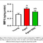 |
Figure 2: Effect of blueberry extract on renal level of matrix metallopeptidase 9 (MMP-9) in aged rats. |
Effect of blueberry extract on renal levels of oxidative stress markers
In the aged rat group, an elevation in the levels of malondialdehyde (MDA; p < 0.001 vs control rats), a biomarker used to assess lipid peroxidation and oxidative damage 37, as well as nitric oxide (NO; p < 0.001 vs control rats) was observed (Figure 3). Furthermore, the same group, exhibited a notable decrease in the levels of reduced glutathione (GSH; p < 0.001 vs control rats), a crucial antioxidant molecule responsible for mitigating oxidative stress, as well as a loss in the total antioxidant capacity (TAOC; p < 0.001 vs control rats) of the renal tissues.
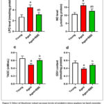 |
Figure 3: Effect of blueberry extract on renal levels of oxidative stress markers (a) lipid peroxidation, (b) nitric oxide, (c) glutathione, and (d) total antioxidant capacity in aged rats. |
Nevertheless, by administering BBE to the aged group, significant alterations in the oxidative stress indicators were detected. The use of BBE led to a significant decrease in the concentrations of MDA (p < 0.001 vs aged rats) and NO (p < 0.001 vs aged rats) in the renal tissues. In addition, the administration of BBE resulted in an augmentation of GSH levels (p < 0.002 vs aged rats), which plays a crucial role in protecting against oxidative harm, alongside an elevation in the overall antioxidant capacity (TAOC; p < 0.002 vs aged rats) of the renal tissues. According to these results, BBE may help protect renal tissues from oxidative damage and enhance their antioxidant defense systems as they age.
Effect of blueberry extract on renal activity of superoxide dismutase, catalase, glutathione peroxidase, and glutathione reductase
Figure 4 illustrates the significant decreases (p < 0.001 vs control rats) in the enzymatic activity of glutathione reductase, catalase, superoxide dismutase, and glutathione peroxidase in the old group of rats relative to the young group. The observed declines in these antioxidant enzymes point to a diminished capacity of these enzymes to protect the kidneys from oxidative stress as they age.
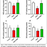 |
Figure 4: Effect of blueberry extract on renal activities of antioxidant enzymes (a) superoxide dismutase, (b) catalase, (c) glutathione peroxidase, and (d) glutathione reductase in aged rats. |
In contrast, the implementation of BBE supplementation among the group of aged rats resulted in notable antioxidant outcomes. The observed phenomenon was evident through significant statistical increases (p < 0.02 vs aged rats) in the concentrations of SOD, GPx, and GR, as compared to the control group. The observed increase in antioxidant enzyme levels indicates that the administration of BBE supplements effectively enhanced the antioxidant defense system in the renal tissues of older rats. This may have the potential to mitigate the increased oxidative stress commonly associated with the aging process.
Moreover, when compared to the control group, a significant improvement (with statistical significance at p < 0.05) in renal CAT activity was noted. The observed increase in CAT activity suggests an enhanced renal capacity to metabolize hydrogen peroxide, a type of reactive oxygen species that has the potential to cause oxidative harm if not adequately neutralized.
In short, the results shown in Figure 4 show substantial changes in the functioning of crucial antioxidant enzymes in the renal system. The aforementioned changes, which were detected in relation to the aging process and the addition of BBE, emphasize the ability of blue berries to alleviate oxidative stress associated with aging. This is accomplished by increasing the functionality of important antioxidant enzymes, hence enhancing antioxidant defences inside the tissues of aged rats.
Effect of blueberry extract on renal expression of nuclear factor erythroid 2-related factor 2
Figure 5 shows that rats in the aged group had significantly lower Nrf2 expression (p < 0.05) when compared to the young control group. However, supplementation with BBE showed a remarkable increase (p < 0.02 vs aged rats) in Nrf2 expression. This decrease in Nrf2 expression suggests a potential decline in the activation of this crucial transcription factor, which plays a pivotal role in orchestrating antioxidant and cytoprotective responses with age. The observed rise in levels suggests that BBE has the ability to effectively modulate the activation of Nrf2, hence potentially augmenting the cellular antioxidant and protective pathways.
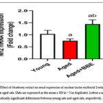 |
Figure 5: Effect of blueberry extract on renal expression of nuclear factor erythroid 2-related factor 2 (Nrf2) in aged rats. |
Effect of blueberry extract on renal levels of inflammatory markers (tumor necrosis factor-α, interleukin-1β, and interleukin-10)
Comparing the aged rat group to the young control group, Figure 6 demonstrates significant increases (p < 0.001 vs control rats) in kidney levels of the inflammatory markers TNF-α and IL-1β. In contrast to the elderly group, BBE supplementation was able to effectively reduce (p < 0.001 vs aged rats) the levels of both inflammatory indicators in the renal tissue. Conversely, the elderly group’s IL-10 level somewhat decreased (p > 0.05 vs control rats); but, after being given blueberries, the group’s IL-10 level significantly increased (p < 0.001 vs aged rats).
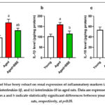 |
Figure 6: Effect of blue berry extract on renal expression of inflammatory markers (a) tumor necrosis factor-α, (b) interleukin-1β, and (c) interleukin-10 in aged rats. |
Histopathological findings in kidney tissues
The histopathological findings in the kidney tissues highlight important structural changes in the kidneys of rats as a consequence of aging and the potential effects of BBE administration. In the kidney sections of young rats, histopathological studies revealed a normal structure. Specifically, the glomeruli (the filtration units of the kidney) and renal tubules (responsible for reabsorbing and excreting substances) in the cortex region appeared to be in their typical state with no significant structural abnormalities (Figure 7A). However, in the aged group, the kidney sections exhibited various pathological alterations, such as congested glomeruli, which suggest compromised blood circulation (Figure 7B and Table1).
In kidney sections of rats treated with BBE, some positive effects were observed. Glomerular structures with well-established epithelia, resembling the control section from young rats, suggest that BBE treatment helped preserve the integrity of the filtration units. However, there were still some mild organized tubules and evidence of atrophy, indicating that while BBE may have had a protective effect, it may not have completely reversed all age-related structural changes in the kidneys (Figure 7C and Table1).
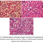 |
Figure 7: A; Photomicrographs of histological changes in the kidney tissue following treatment with blueberry extract in aged rats. |
Blue arrows indicate inflammatory cells, blue stars indicate debris within the lumen of some renal tubules, red stars indicate injured renal tubules, and black arrows indicate pyknosis and karyolysis.
Table 1: Histopathological findings in kidney of aged rats treated with blueberry extract
| Treatment | |||
| Finding | Young | Untrated Aged |
Aged+BBE |
|
Hypertrophy of epithelial cells of renal tubules |
± | ++++ | ++ |
| Degeneration of tubular epithelia with simultaneous infiltration of mononuclear cells | + | +++ |
++ |
|
Hyperaemia of medullary and cortical part with mononuclear cell infiltration |
+ | +++ | ++ |
| Dilation of renal glomeruli | + | ++++ |
++ |
++++, the change was very often found in all animals; +++, the change was relatively common in all animals; ++, the change was rare in all animals; +, the change was found in a few animals; ±, the change was sporadic.
Discussion
The aging kidney has drawn a lot of attention in clinical nephrology and geriatric medicine recently. According to this study, taking supplements of blueberries may be essential for reducing kidney aging. Furthermore, this is the first study that, to the best of our knowledge, focuses on the many processes by which BBE influences the alterations in the aging kidney.
The renal function of aged rats was shown to be impaired, which is in line with the prior reports.38, 39 This impairment was attributed to structural tubular alterations and impairment of the glomerular filtration rate, as suggested by Salem and Faried.40 This was consistent with the study’s findings, which were supported by the older group’s much higher levels of urea and creatinine than those of the younger group. Furthermore, serum creatinine and urea values are usually high due to an augment in the habitual creatinine back-filtration in the aged 39. The decrease in blood calcium levels could have implications for various physiological processes, potentially reflecting age-related changes in calcium regulation and metabolism.41
However, administering BBE in the Aged+BBE group extensively declined the levels of serum urea (p < 0.03 vs aged rats) and creatinine (p < 0.001 vs aged rats) while restoring calcium levels (p < 0.001 vs aged rats) back to normal. The preventive properties of BBE could be linked to its ameliorative impact on metabolic alterations that accompany aging, as well as its antioxidant properties. Accordingly, blueberries have a high concentration of active phenolics and flavonoids that may be able to reduce fibrosis, oxidative stress, and inflammation. Pterostilbene, a dimethyl ether counterpart of resveratrol found naturally in blueberries, demonstrated anti-hyperuricemic action and improved kidney functioning in rats administered potassium oxonate to treat mild hyperuricemia in earlier research.42 Unfortunately, Huang 43 found that the long-term L-arginine supplementation did not show any benefit effect on kidney. It rather accelerates functional decline of kidney and vasculature in aging. Thus, the long-term dietary L-arginine supplementation should be avoided particularly in elderly population.
The study’s findings showed that the older rats had higher MMP-9 levels whereas, BBE administration restrained it. An enzyme called MMP-9 is involved in the breakdown of extracellular matrix components, which is necessary for tissue remodelling and healing.44 The human kidney’s mesangial cells, tubular epithelial cells, etc., can normally manufacture MMP-2 and 9, although only in small amounts. On the other hand, aberrant activation and interactions of numerous cells signalling pathways cause the mRNA transcription levels of MMP-2 and 9 to be rapidly increased during the course of renal fibrosis 45. The potential reason for the increase in MMP-9 could be attributed to TNF-α capacity to stimulate the production of MMP-2 and 9, as well as to encourage the activation of pro-MMP-2, which in turn increases MMP-2 activity. TNF-α has the ability to stimulate pro-MMP-9 activation as well. On the other hand, the conversion of pro-TNF-α to TNF-α will likewise be prevented if MMP-2 and 9 activities are decreased 46. Additionally, as was previously indicated, oxidative stress can activate MMP-2 and 9, the activities of which can vary depending on the stage of chronic kidney disease 47. Renal interstitial fibrosis deteriorates more with the course of CKD, impeding oxygen diffusion and ultimately resulting in severe hypoxia. The hypoxia exacerbates OS even more, starting a vicious cycle that ultimately intensifies fibrosis.45
This observation suggests that the administration of BBE could potentially have a modulatory effect on MMP-9 levels in aged rats’ kidneys, indicating a potential beneficial impact on the enzymatic environment within the renal tissues of the aging population.
As was already indicated, the general findings of this investigation are consistent with those of numerous prior studies48-50, which indicate that the activity of CAT in the liver and renal tissues of mice and rats decreases with age. Furthermore, the results of this study broadly concur with those of previous investigations 51 showing that glutathione utilizing and recycling system activity in the kidney also decline with advancing age. Moreover, the observed decline in SOD activity with age is consistent with previous observations.52 Overall, a wealth of research links aging to a decline in antioxidant status.48, 53 Oxidative stress is one of the possible inducers of senescence, since ROS production can damage the DNA and other macromolecules.54
The SOD family of intracellular enzymes, which catalyses dismutation, prevents damage to biological structures and membranes from oxygen free radicals by catalysing the elimination of a superoxide radical (O·−2). Significant amounts of H2O2 have been demonstrated to be detoxified by CAT.55 The effectiveness of mitochondrial defence systems against the disorganizing effects of free radicals is weakened as SOD activity declines. The findings reported here are consistent with a previous study by Subramanian and James52, which found that SOD activity somewhat decreased with aging. However, the current study expands on this research by offering a more thorough understanding of age-related changes, emphasising the coordinated response of the entire spectrum of antioxidant enzymes and critically evaluating the significance of the regional distribution of the enzyme activities.
Aging impairs antioxidant balance as demonstrated by Marquez-Exposito 56 who found that 18-month-old mice has impairment nuclear factor (erythroid-derived 2)-related factor 2 pathway, as evidenced by the downregulation of active Nrf2 and decreased antioxidant enzyme gene expression. The fact that Nrf2 gene deletion increased renal oxidative stress and inflammation in the animal model of diabetes 57 supports the protective function of Nrf2 on the kidneys. Furthermore, compared to wild-type mice, Nrf2 knockout animals showed more severe kidney damage under ischemia and nephrotoxic insults.58 Conversely, in a number of experimental models, pharmaceutical therapies employing Nrf2 activators reduced markers of oxidative stress-induced kidney injury.59
Furthermore, under oxidative stress circumstances, the reduction of Nrf2 antioxidant capacity exacerbates renal tubular apoptosis and atrophy as well as interstitial fibrosis.60 The present investigation revealed that the administration of BBE reversed the downregulation of Nrf2 and the oxidative stress that was subsequently caused by aging in the kidney tissues. This suggests that BBE may have an effect on activating the Nrf2 signalling pathway in kidneys that are older. In this regard, a recent study found that by upregulating Nrf2, ellagic acid could significantly protect kidneys from oxidative stress caused by aging-induced renal oxidative damage through upregulating SIRT1 and Nrf2.61 Furthermore, it has been established that blueberry polyphenols prevents angiotensin II-induced oxidative stress and inflammatory signaling in human aortic endothelial cells via modifying Nrf2 transcription and activity.62
The obtained results are similar to the results of Pokkunuri 63 who found that grape powder potentially regulating Nrf2 that improved mitochondrial and kidney functions in aged Fischer 344 rats. Moreover, the ability of natural products to improve antioxidant defence system in aged rats was investigated largely by other researchers. In this regard, Samarghandian 64 found that Crocus sativus L. (saffron) extract improved the antioxidant system and inhibited the inflammation response in the kidney of old rats and with Farzanegi 65 who reported that garlic extract not only improved antioxidant capacity but also declined oxidative damages and apoptosis through reducing Bax levels and enhancing Bcl-2 value in liver and kidney tissues of aged rats.
The ageing process can be responsible for the observed increases in kidney levels of the inflammatory markers’ tumour necrosis factor-alpha and interleukin-1 beta in the old group as compared to the young control group. The process of aging is frequently linked to the presence of a persistent, low-grade inflammation known as “inflammaging”.66 This condition is characterized by an impaired immune system that exhibits dysregulation and an elevated production of pro-inflammatory cytokines such as TNF-α and IL-1β. 66, 67 Age-related inflammation can exert negative effects on tissue function, specifically impacting the kidneys.68 The observed decrease in TNF-α and IL-1β levels following the treatment of blueberries in the aged rats suggests that blueberries possess anti-inflammatory effects. Blueberries are abundant in bioactive chemicals, namely anthocyanins, and flavonoids, that have demonstrated anti-inflammatory benefits.11 These chemicals have the ability to regulate inflammatory pathways, leading to a decrease in the synthesis of pro-inflammatory cytokines such as TNF-α and IL-1β.43
Conversely, a slight decline in Interleukin-10 levels within the aged group was observed. IL-10 is an anti-inflammatory cytokine known to dampen inflammatory responses.69 The immune system undergoes age-related modifications that might lead to changes in the synthesis and control of anti-inflammatory cytokines such as IL-10.70 Remarkably, subsequent to the administration of BBE, a significant elevation in IL-10 levels was observed. This finding implies that blueberries might have a modulatory impact on the immune response, facilitating the development of an anti-inflammatory environment in the kidneys of aged rats. These findings suggest BBE administration appears to mitigate these age-related effects by reducing pro-inflammatory markers and boosting anti-inflammatory cytokines, contributing to a more balanced and less inflammatory renal environment in aged rats.
Several studies have shown that blueberry components can lower proinflammatory marker gene expression and secretion in distinct cell models (RAW 264.7 and bone marrow-derived macrophage (BMDMs)]. In RAW 264.7 cells and BMDMs, blueberry phytochemicals decreased gene expression and secretion of proinflammatory cytokines generated by lipopolysaccharides (LPS), specifically IL-6 71, IL-1β 72, and TNF-α.73 Furthermore, blueberry phytochemicals have been shown to have anti-inflammatory properties in human cell lines 74, including the U-937 and THP-1 monocyte-like cells, which can be differentiated into macrophages after stimulation with phorbol-12-myristate-13-acetate (PMA) 75, and human primary peripheral blood mononuclear cells (PBMCs). Most investigations linked the regulatory impact to a decrease in NF-κB translocation in the human monocytic leukemia cell line THP-1.76
The presence of inflammatory cells suggests an inflammatory response, which may indicate renal impairment. The presence of debris within the lumen of some renal tubules indicates a potential impairment of tubular function.77, 78 Also, the presence of injured renal tubules with vacuolated cytoplasm implies structural impairment to these essential constituents of the renal system.79, 80 The presence of pyknosis (shrinkage and condensation of the cell nucleus) and karyolysis (dissolution of the cell nucleus) in some tubular cells suggests cellular damage and potential cell death, which are concerning signs of kidney injury or aging-related changes.81, 82 However, the ability of BBE to protect renal cells from damage is consistent with the results of Nair 83 who found that BB protects against acute kidney injury by inhibiting Toll-like receptor 4 (TLR4) and its subsequent effect on inflammatory and oxidative stress pathways. The same was demonstrated by Elks 84 who reported the nephroprotective effect of blueberries in a rat model of hypertension.
Study limitations
Several limitations should be taken into account, even if this study offers insightful information about the possible advantages of blueberry extract (BBE) supplementation on age-related renal abnormalities. The results may not be as broadly applicable to humans due to the small sample size and animal model. Furthermore, the study’s narrow age range and emphasis on male rats might have missed subtle age-related changes or possible sex-specific effects. Additionally, the study needs to extend to determine which active ingredient in BBE was responsible for this effect and to analysis the crude extract of BB. The study points out that in order to completely clarify the therapeutic potential of BBE in reducing age-related kidney alterations and to ascertain its application in human health contexts, more research addressing these limitations—including human clinical trials—is required.
Conclusion
In summary, the comprehensive results and discussions presented in this study shed light on the potential benefits of BBE supplementation in mitigating age-related renal changes and oxidative stress in rats. The aging process was associated with significant alterations in various renal parameters, including increased levels of serum urea and creatinine, decreased blood calcium levels, elevated MMP-9, oxidative stress, and reduced antioxidant enzyme activities. However, the introduction of BBE in the aged rats exhibited several positive effects, such as lowering serum levels of urea and creatinine, restoring blood calcium levels, reducing MMP-9, alleviating oxidative stress, and enhancing antioxidant defense mechanisms. Additionally, BBE demonstrated anti-inflammatory properties by suppressing the pro-inflammatory markers TNF-α and IL-1β while increasing the levels of the anti-inflammatory cytokine IL-10. Histopathological examination highlighted age-related structural changes in the kidneys, which were partially mitigated by BBE, particularly in preserving glomerular integrity. These findings collectively suggest that BBE may have a therapeutic potential in countering age-related renal oxidative stress and inflammation, emphasizing its role as a dietary intervention for promoting kidney health in aging individuals. Further research, including clinical trials, is warranted to validate these findings and explore their applicability in human contexts. Additionally, further studies on different age groups determine the optimal age for starting BBE supplementation and to determine the optimal dosage for humans based on the rat studies.
Acknowledgement
Princess Nourah bint Abdulrahman University Researchers Supporting Project number (PNURSP2024R69), Princess Nourah bint Abdulrahman University, Riyadh, Saudi Arabia.
Funding Sources
Princess Nourah bint Abdulrahman University Researchers Supporting Project number (PNURSP2024R69), Princess Nourah bint Abdulrahman University, Riyadh, Saudi Arabia.
Conflicts of Interest
The author declares that there is no conflict of interest.
Data Availability Statement
All the data obtained is available within the manuscript.
Ethics Statement
The experimental procedures were performed concomitantly with the National Institutes of Health (NIH) Guidelines for the Care and Use of Laboratory Animals, 8th edition (NIH Publication No. 85-23, revised 1985) and approved by the Princess Nourah bint Abdulrahman University Institutional Animal Care and Use Committee (IACUC) criteria (Approval No. HAP-01-R-059; IRB Registration No.: 22-0161; Category of Approval: EXEMPT). To ensure animal welfare, all in vivo experiments were performed in accordance with the ARRIVE guidelines.
Informed Consent Statement
This study did not involve human participants, and therefore, informed consent was not required.
Permission to Reproduce Material from Other
Not applicable.
Clinical Trial Registration
Not applicable.
Author Contributions
Tarfa Albrahim : Conceptualization, methodology, validation, formal analysis, investigation, resources, data curation, writing—original draft preparation, writing—review and editing, funding acquisition.
References
- Kassis A, Fichot MC, Horcajada MN, et al. Nutritional and lifestyle management of the aging journey: A narrative review. Front Nutr. 2022;9:1087505. doi:10.3389/fnut.2022.1087505
CrossRef - Ray N, Reddy PH. Structural and physiological changes of the kidney with age and its impact on chronic conditions and COVID-19. Ageing Res Rev. Jul 2023;88:101932. doi:10.1016/j.arr.2023.101932
CrossRef - Hashmi MF, Benjamin O, Lappin SL. End-Stage Renal Disease. StatPearls. 2023.
- Denic A, Glassock RJ, Rule AD. Structural and Functional Changes With the Aging Kidney. Adv Chronic Kidney Dis. Jan 2016;23(1):19-28. doi:10.1053/j.ackd.2015.08.004
CrossRef - Noronha IL, Santa-Catharina GP, Andrade L, Coelho VA, Jacob-Filho W, Elias RM. Glomerular filtration in the aging population. Front Med (Lausanne). 2022;9:769329. doi:10.3389/fmed.2022.769329
CrossRef - Bialik VV. [Incidence and morphological characteristics of prostatic cancer depending on patients’ age]. Klin Khir (1962). Oct 1974;(10):64-6. O chastote i morfologicheskikh osobennostiiakh raka predstatel’noi zhelezy v zavisimosti ot vozrasta.
- Ferenbach DA, Bonventre JV. Acute kidney injury and chronic kidney disease: From the laboratory to the clinic. Nephrol Ther. Apr 2016;12 Suppl 1(Suppl 1):S41-8. doi:10.1016/j.nephro.2016.02.005
CrossRef - Fekete M, Szarvas Z, Fazekas-Pongor V, et al. Nutrition Strategies Promoting Healthy Aging: From Improvement of Cardiovascular and Brain Health to Prevention of Age-Associated Diseases. Nutrients. Dec 22 2022;15(1)doi:10.3390/nu15010047
CrossRef - Mousa D, Alharbi A, Helal I, et al. Prevalence and Associated Factors of Chronic Kidney Disease among Relatives of Hemodialysis Patients in Saudi Arabia. Kidney Int Rep. Mar 2021;6(3):817-820. doi:S2468-0249(20)31863-5 [pii] 10.1016/j.ekir.2020.12.029
CrossRef - Alsuwaida AO, Farag YM, Al Sayyari AA, et al. Epidemiology of chronic kidney disease in the Kingdom of Saudi Arabia (SEEK-Saudi investigators) – a pilot study. Saudi J Kidney Dis Transpl. Nov 2010;21(6):1066-72. doi:SaudiJKidneyDisTranspl_2010_21_6_1066 _72293 [pii]
- Kalt W, Cassidy A, Howard LR, et al. Recent Research on the Health Benefits of Blueberries and Their Anthocyanins. Adv Nutr. Mar 1 2020;11(2):224-236. doi:5536953 [pii] 10.1093/advances/nmz065
CrossRef - Albrahim T, Alonazi M. Effect of Blueberry Extract on Liver in Aged Rats. Oxid Med Cell Longev. 2022;2022:3490776. doi:10.1155/2022/3490776
CrossRef - Felgus-Lavefve L, Howard L, Adams SH, Baum JI. The Effects of Blueberry Phytochemicals on Cell Models of Inflammation and Oxidative Stress. Adv Nutr. Aug 1 2022;13(4):1279-1309. doi:10.1093/advances/nmab137
CrossRef - Song G-Q, Hancock JF. Recent Advances in Blueberry Transformation. doi: 10.1080/15538362.2011.623083. International Journal of Fruit Science. 2012/01/01 2012;12(1-3):316-332. doi:10.1080/15538362.2011.623083
CrossRef - Krishna P, Pandey G, Thomas R, Parks S. Improving Blueberry Fruit Nutritional Quality through Physiological and Genetic Interventions: A Review of Current Research and Future Directions. Antioxidants. 2023;12. doi:10.3390/antiox12040810
CrossRef - Nadulski R, Masłowski A, Mazurek A, et al. Vitamin C and lutein content of northern highbush blueberry (Vaccinium corymbosum L.) juice processed using freezing and thawing. Journal of Food Measurement and Characterization. 2019/12/01 2019;13(4):2521-2528. doi:10.1007/s11694-019-00172-x
CrossRef - Drozdz P, Seziene V, Pyrzynska K. Mineral Composition of Wild and Cultivated Blueberries. Biol Trace Elem Res. Jan 2018;181(1):173-177. doi:10.1007/s12011-017-1033-z 1033 [pii] 10.1007/s12011-017-1033-z
CrossRef - Tran PHL, Tran TTD. Blueberry Supplementation in Neuronal Health and Protective Technologies for Efficient Delivery of Blueberry Anthocyanins. Biomolecules. Jan 14 2021;11(1)doi:biom11010102 [pii] biomolecules-11-00102 [pii] 10.3390/biom11010102
CrossRef - Grosso G, Micek A, Godos J, et al. Dietary Flavonoid and Lignan Intake and Mortality in Prospective Cohort Studies: Systematic Review and Dose-Response Meta-Analysis. Am J Epidemiol. Jun 15 2017;185(12):1304-1316. doi:10.1093/aje/kww207
CrossRef - Wang X, Ouyang YY, Liu J, Zhao G. Flavonoid intake and risk of CVD: a systematic review and meta-analysis of prospective cohort studies. Br J Nutr. Jan 14 2014;111(1):1-11. doi:10.1017/S000711451300278X
CrossRef - Cassidy A, Mukamal KJ, Liu L, Franz M, Eliassen AH, Rimm EB. High anthocyanin intake is associated with a reduced risk of myocardial infarction in young and middle-aged women. Circulation. Jan 15 2013;127(2):188-96. doi:10.1161/CIRCULATIONAHA .112.122408
CrossRef - Ellis KJ. Human body composition: in vivo methods. Physiological reviews. Apr 2000;80(2):649-80. doi:10.1152/physrev.2000.80.2.649
CrossRef - Jennings A, MacGregor A, Spector T, Cassidy A. Higher dietary flavonoid intakes are associated with lower objectively measured body composition in women: evidence from discordant monozygotic twins. Am J Clin Nutr. Mar 2017;105(3):626-634. doi:10.3945/ajcn.116.144394
CrossRef - Muraki I, Imamura F, Manson JE, et al. Fruit consumption and risk of type 2 diabetes: results from three prospective longitudinal cohort studies. BMJ. Aug 28 2013;347:f5001. doi:10.1136/bmj.f5001
CrossRef - Wedick NM, Pan A, Cassidy A, et al. Dietary flavonoid intakes and risk of type 2 diabetes in US men and women. Am J Clin Nutr. Apr 2012;95(4):925-33. doi:10.3945/ajcn.111.028894
CrossRef - McNamara RK, Kalt W, Shidler MD, et al. Cognitive response to fish oil, blueberry, and combined supplementation in older adults with subjective cognitive impairment. Neurobiol Aging. Apr 2018;64:147-156. doi:10.1016/j.neurobiolaging.2017.12.003
CrossRef - Li X, Zhu F, Zeng Z. Effects of different extraction methods on antioxidant properties of blueberry anthocyanins. Open Chemistry. 2021;19(1):138-148. Open Chemistry. doi:doi:10.1515/chem-2020-0052
CrossRef - Debom G, Gazal M, Soares MS, et al. Preventive effects of blueberry extract on behavioral and biochemical dysfunctions in rats submitted to a model of manic behavior induced by ketamine. Brain research bulletin. Oct 2016;127:260-269. doi:S0361-9230(16)30352-5 [pii]10.1016/j.brainresbull.2016.10.008
CrossRef - Ohkawa H, Ohishi N, Yagi K. Assay for Lipid Peroxides in Animal-Tissues by Thiobarbituric Acid Reaction. Anal Biochem. 1979;95(2):351-358. doi:Doi 10.1016/0003-2697(79)90738-3
CrossRef - Green LC, Wagner DA, Glogowski J, Skipper PL, Wishnok JS, Tannenbaum SR. Analysis of nitrate, nitrite, and [15N]nitrate in biological fluids. Anal Biochem. Oct 1982;126(1):131-8. doi:0003-2697(82)90118-X [pii]
CrossRef - Rubio CP, Hernandez-Ruiz J, Martinez-Subiela S, Tvarijonaviciute A, Ceron JJ. Spectrophotometric assays for total antioxidant capacity (TAC) in dog serum: an update. BMC Vet Res. Aug 15 2016;12(1):166. doi:10.1186/s12917-016-0792-7
CrossRef - Ellman GL. Tissue Sulfhydryl Groups. Arch Biochem Biophys. 1959;82(1):70-77. doi:Doi 10.1016/0003-9861(59)90090-6
CrossRef - Nishikimi M, Appaji N, Yagi K. The occurrence of superoxide anion in the reaction of reduced phenazine methosulfate and molecular oxygen. Biochem Biophys Res Commun. Jan 31 1972;46(2):849-54.
CrossRef - Aebi H. Catalase in vitro. Methods Enzymol. 1984;105:121-6.
CrossRef - De Vega L, Fernandez RP, Mateo MC, Bustamante JB, Herrero AM, Munguira EB. Glutathione determination and a study of the activity of glutathione-peroxidase, glutathione-transferase, and glutathione-reductase in renal transplants. Ren Fail. Jul 2002;24(4):421-32.
CrossRef - Paglia DE, Valentine WN. Studies on the quantitative and qualitative characterization of erythrocyte glutathione peroxidase. J Lab Clin Med. Jul 1967;70(1):158-69. doi:0022-2143(67)90076-5 [pii]
- Tsikas D. Assessment of lipid peroxidation by measuring malondialdehyde (MDA) and relatives in biological samples: Analytical and biological challenges. Anal Biochem. May 1 2017;524:13-30. doi:10.1016/j.ab.2016.10.021
CrossRef - Costa E, Fernandes J, Ribeiro S, et al. Aging is Associated with Impaired Renal Function, INF-gamma Induced Inflammation and with Alterations in Iron Regulatory Proteins Gene Expression. Aging Dis. Dec 2014;5(6):356-65. doi:ad-5-6-356 [pii] 10.14366/ AD.2014.0500356
- Musso CG, Alvarez Gregori J, Jauregui JR, Macias Nunez JF. Creatinine, urea, uric acid, water and electrolytes renal handling in the healthy oldest old. World J Nephrol. Oct 6 2012;1(5):123-6. doi:10.5527/wjn.v1.i5.123
CrossRef - Salem HR, Faried MA. Treadmill Exercise Training Ameliorates Functional and Structural Age-Associated Kidney Changes in Male Albino Rats. ScientificWorldJournal. 2021;2021:1393372. doi:10.1155/2021/1393372
CrossRef - Veldurthy V, Wei R, Oz L, Dhawan P, Jeon YH, Christakos S. Vitamin D, calcium homeostasis and aging. Bone Res. 2016;4:16041. doi:10.1038/boneres.2016.41
CrossRef - Pan J, Shi M, Li L, et al. Pterostilbene, a bioactive component of blueberries, alleviates renal fibrosis in a severe mouse model of hyperuricemic nephropathy. Biomed Pharmacother. Jan 2019;109:1802-1808. doi:S0753-3322(18)35726-3 [pii] 10.1016/j.biopha.2018.11.022
CrossRef - Huang W, Yan Z, Li D, Ma Y, Zhou J, Sui Z. Antioxidant and Anti-Inflammatory Effects of Blueberry Anthocyanins on High Glucose-Induced Human Retinal Capillary Endothelial Cells. Oxid Med Cell Longev. 2018;2018:1862462. doi:10.1155/2018/1862462
CrossRef - Cabral-Pacheco GA, Garza-Veloz I, Castruita-De la Rosa C, et al. The Roles of Matrix Metalloproteinases and Their Inhibitors in Human Diseases. Int J Mol Sci. Dec 20 2020;21(24)doi:10.3390/ijms21249739
CrossRef - Cheng Z, Limbu MH, Wang Z, et al. MMP-2 and 9 in Chronic Kidney Disease. Int J Mol Sci. Apr 8 2017;18(4)doi:ijms18040776 [pii] ijms-18-00776 [pii] 10.3390/ijms18040776
CrossRef - Gearing AJ, Beckett P, Christodoulou M, et al. Matrix metalloproteinases and processing of pro-TNF-alpha. Journal of leukocyte biology. May 1995;57(5):774-7. doi:10.1002/jlb.57.5.774
CrossRef - Pulido-Olmo H, Garcia-Prieto CF, Alvarez-Llamas G, et al. Role of matrix metalloproteinase-9 in chronic kidney disease: a new biomarker of resistant albuminuria. Clin Sci (Lond). Apr 1 2016;130(7):525-38. doi:CS20150517 [pii] 10.1042/CS20150517
CrossRef - Thiab NR, King N, Jones GL. Effect of ageing and oxidative stress on antioxidant enzyme activity in different regions of the rat kidney. Mol Cell Biochem. Oct 2015;408(1-2):253-60. doi:10.1007/s11010-015-2503-2 [pii] 10.1007/s11010-015-2503-2
CrossRef - Carney Almroth B, Johnsson JI, Devlin R, Sturve J. Oxidative stress in growth hormone transgenic coho salmon with compressed lifespan–a model for addressing aging. Free radical research. Oct 2012;46(10):1183-9. doi:10.3109/10715762.2012.698009
CrossRef - Farahmand SK, Samini F, Samini M, Samarghandian S. Safranal ameliorates antioxidant enzymes and suppresses lipid peroxidation and nitric oxide formation in aged male rat liver. Biogerontology. Feb 2013;14(1):63-71. doi:10.1007/s10522-012-9409-0
CrossRef - Ramesh T, Kim SW, Sung JH, et al. Effect of fermented Panax ginseng extract (GINST) on oxidative stress and antioxidant activities in major organs of aged rats. Experimental gerontology. Jan 2012;47(1):77-84. doi:S0531-5565(11)00304-4 [pii] 10.1016/j.exger.2011. 10.007
CrossRef - Subramanian MV, James TJ. Age-related protective effect of deprenyl on changes in the levels of diagnostic marker enzymes and antioxidant defense enzymes activities in cerebellar tissue in Wistar rats. Cell stress & chaperones. Sep 2010;15(5):743-51. doi:177 [pii] 10.1007/s12192-010-0177-y
CrossRef - Shih PH, Yen GC. Differential expressions of antioxidant status in aging rats: the role of transcriptional factor Nrf2 and MAPK signaling pathway. Biogerontology. Apr 2007;8(2):71-80. doi:10.1007/s10522-006-9033-y
CrossRef - Abdel Moneim AE. Indigofera oblongifolia prevents lead acetate-induced hepatotoxicity, oxidative stress, fibrosis and apoptosis in rats. PLoS One. 2016;11(7):e0158965.
CrossRef - Almeer RS, AlBasher GI, Alarifi S, Alkahtani S, Ali D, Abdel Moneim AE. Royal jelly attenuates cadmium-induced nephrotoxicity in male mice. Sci Rep. Apr 9 2019;9(1):5825. doi:10.1038/s41598-019-42368-7 10.1038/s41598-019-42368-7
CrossRef - Marquez-Exposito L, Tejedor-Santamaria L, Valentijn FA, et al. Oxidative Stress and Cellular Senescence Are Involved in the Aging Kidney. Antioxidants (Basel). Jan 31 2022;11(2)doi:antiox11020301 antioxidants-11-0030110.3390/antiox11020301
CrossRef - Ruiz S, Pergola PE, Zager RA, Vaziri ND. Targeting the transcription factor Nrf2 to ameliorate oxidative stress and inflammation in chronic kidney disease. Kidney Int. Jun 2013;83(6):1029-41. doi:S0085-2538(15)55858-410.1038/ki.2012.439
CrossRef - Liu M, Grigoryev DN, Crow MT, et al. Transcription factor Nrf2 is protective during ischemic and nephrotoxic acute kidney injury in mice. Kidney Int. Aug 2009;76(3):277-85. doi:S0085-2538(15)53959-8 [pii]10.1038/ki.2009.157
CrossRef - Choi BH, Kang KS, Kwak MK. Effect of redox modulating NRF2 activators on chronic kidney disease. Molecules. Aug 20 2014;19(8):12727-59. doi:molecules190812727 [pii]molecules-19-12727 [pii]10.3390/molecules190812727
CrossRef - Wu CF, Liu CC, Tsai YC, Chen CC, Wu MT, Hsieh TJ. Diminishment of Nrf2 Antioxidative Defense Aggravates Nephrotoxicity of Melamine and Oxalate Coexposure. Antioxidants (Basel). Sep 14 2021;10(9)doi:antiox10091464 [pii]antioxidants-10-01464 [pii] 10.3390/antiox10091464
CrossRef - Naghibi N, Sadeghi A, Movahedinia S, et al. Ellagic acid ameliorates aging-induced renal oxidative damage through upregulating SIRT1 and NRF2. BMC complementary medicine and therapies. 2023/03/10 2023;23(1):77. doi:10.1186/s12906-023-03907-y
CrossRef - Najjar RS, Mu S, Feresin RG. Blueberry Polyphenols Increase Nitric Oxide and Attenuate Angiotensin II-Induced Oxidative Stress and Inflammatory Signaling in Human Aortic Endothelial Cells. Antioxidants (Basel). Mar 23 2022;11(4)doi:antiox11040616 [pii] antioxidants-11-00616 [pii] 10.3390/antiox11040616
CrossRef - Pokkunuri I, Ali Q, Asghar M. Grape Powder Improves Age-Related Decline in Mitochondrial and Kidney Functions in Fischer 344 Rats. Oxidative medicine and cellular longevity. 2016/07/27 2016;2016:6135319. doi:10.1155/2016/6135319
CrossRef - Samarghandian S, Azimi-Nezhad M, Borji A, Farkhondeh T. Crocus sativus L. (saffron) extract reduces the extent of oxidative stress and proinflammatory state in aged rat kidney. Progress in Nutrition. 2016;18(3):299-310.
- Farzanegi P, Abbaszadeh H, Farokhi F, et al. Attenuated Renal and Hepatic Cells Apoptosis Following Swimming Exercise Supplemented with Garlic Extract in Old Rats. Clin Interv Aging. 2020;15:1409-1418. doi:250321 [pii] 10.2147/CIA.S250321
CrossRef - Ferrucci L, Fabbri E. Inflammageing: chronic inflammation in ageing, cardiovascular disease, and frailty. Nat Rev Cardiol. Sep 2018;15(9):505-522. doi:10.1038/s41569-018-0064-2
CrossRef - Zuo L, Prather ER, Stetskiv M, et al. Inflammaging and Oxidative Stress in Human Diseases: From Molecular Mechanisms to Novel Treatments. Int J Mol Sci. Sep 10 2019;20(18)doi:10.3390/ijms20184472
CrossRef - Ebert T, Pawelzik SC, Witasp A, et al. Inflammation and Premature Ageing in Chronic Kidney Disease. Toxins (Basel). Apr 4 2020;12(4)doi:10.3390/toxins12040227
CrossRef - Steen EH, Wang X, Balaji S, Butte MJ, Bollyky PL, Keswani SG. The Role of the Anti-Inflammatory Cytokine Interleukin-10 in Tissue Fibrosis. Adv Wound Care (New Rochelle). Apr 1 2020;9(4):184-198. doi:10.1089/wound.2019.1032
CrossRef - Rea IM, Gibson DS, McGilligan V, McNerlan SE, Alexander HD, Ross OA. Age and Age-Related Diseases: Role of Inflammation Triggers and Cytokines. Front Immunol. 2018;9:586. doi:10.3389/fimmu.2018.00586
CrossRef - Esposito D, Chen A, Grace MH, Komarnytsky S, Lila MA. Inhibitory effects of wild blueberry anthocyanins and other flavonoids on biomarkers of acute and chronic inflammation in vitro. J Agric Food Chem. Jul 23 2014;62(29):7022-8. doi:10.1021/jf4051599
CrossRef - Grace MH, Esposito D, Dunlap KL, Lila MA. Comparative analysis of phenolic content and profile, antioxidant capacity, and anti-inflammatory bioactivity in wild Alaskan and commercial Vaccinium berries. J Agric Food Chem. May 7 2014;62(18):4007-17. doi:10.1021/jf403810y
CrossRef - Lee SG, Kim B, Yang Y, et al. Berry anthocyanins suppress the expression and secretion of proinflammatory mediators in macrophages by inhibiting nuclear translocation of NF-kappaB independent of NRF2-mediated mechanism. The Journal of nutritional biochemistry. Apr 2014;25(4):404-11. doi:S0955-2863(13)00261-1 [pii]10.1016/j.jnutbio.2013.12.001
CrossRef - Chanput W, Mes JJ, Wichers HJ. THP-1 cell line: an in vitro cell model for immune modulation approach. Int Immunopharmacol. Nov 2014;23(1):37-45. doi:S1567-5769(14)00316-6 [pii] 10.1016/j.intimp.2014.08.002
CrossRef - Zeng C, Wang W, Yu X, Yang L, Chen S, Li Y. Pathways related to PMA-differentiated THP1 human monocytic leukemia cells revealed by RNA-Seq. Sci China Life Sci. Dec 2015;58(12):1282-7. doi:10.1007/s11427-015-4967-4 [pii]10.1007/s11427-015-4967-4
CrossRef - Kunrade L, Rembergs R, Jēkabsons K, et al. Inhibition of NF- κB pathway in LPS-stimulated THP-1 monocytes and COX-2 actiity in vitro by berry pomace extracts from five Vaccinium species. Journal of Berry Research. 2020;10(3):381-396. doi:10.3233/jbr-190485
CrossRef - Basile DP, Anderson MD, Sutton TA. Pathophysiology of acute kidney injury. Compr Physiol. Apr 2012;2(2):1303-53. doi:10.1002/cphy.c110041
CrossRef - Othman MS, Khaled AM, Aleid GM, et al. Evaluation of antiobesity and hepatorenal protective activities of Salvia officinalis extracts pre-treatment in high-fat diet-induced obese rats. Environmental science and pollution research international. Jun 1 2022;doi:10.1007/s11356-022-21092-2 10.1007/s11356-022-21092-2.
CrossRef - Gaut JP, Liapis H. Acute kidney injury pathology and pathophysiology: a retrospective review. Clin Kidney J. Feb 2021;14(2):526-536. doi:10.1093/ckj/sfaa142
CrossRef - Kaya Y, Bas O, Hanci H, et al. Acute renal involvement in organophosphate poisoning: histological and immunochemical investigations. Ren Fail. Nov 2018;40(1):410-415. doi:10.1080/0886022X.2018.1489289
CrossRef - Elmore SA, Dixon D, Hailey JR, et al. Recommendations from the INHAND Apoptosis/Necrosis Working Group. Toxicol Pathol. Feb 2016;44(2):173-88. doi:10.1177/0192623315625859
CrossRef - Najdawi ZR, Abu-Asab MS. An Ultrastructural Perspective on Cell Death. Jordan Med J. 2022;56(1)doi:10.35516/jmj.v56i1.232
CrossRef - Nair AR, Masson GS, Ebenezer PJ, Del Piero F, Francis J. Role of TLR4 in lipopolysaccharide-induced acute kidney injury: Protection by blueberry. Free Radical Biology and Medicine. 2014/06/01/ 2014;71:16-25. doi:https://doi.org/10.1016/j.freeradbiomed. 2014.03.012
CrossRef - Elks CM, Reed SD, Mariappan N, et al. A blueberry-enriched diet attenuates nephropathy in a rat model of hypertension via reduction in oxidative stress. PLOS ONE. 2011;6(9):e24028. doi:PONE-D-11-06669 [pii] 10.1371/journal.pone.0024028
CrossRef

This work is licensed under a Creative Commons Attribution 4.0 International License.


