
Introduction
The World Health Organization defines health as a state of complete physical, mental, and social well-being rather than only the absence of illness or incapacity. Generally, maintaining good health requires a balanced diet, regular exercise, and enough sleep. Due to risk factors like depression1–5 and anxiety6–7 lifestyle disorders such as obesity, hypertension, cardiovascular disease, and diabetes have recently shown high prevalence. According to the 2019–20 National Family Health Survey, 24% of men and 21% of women had hypertension. Uncontrolled hypertension contributes to various cardiovascular issues, including thrombosis.8 Many factors, including lifestyle, diet, environmental factors, and/or genetics, influence abnormal coagulation and may lead to thrombosis. Currently, many medications are available to treat thrombosis and dissolve blood clots, but the main drawbacks of these drugs are overbleeding, organ damage and hypersensitivity etc.9-10 In the present scenario, research on natural products and their derivatives is gaining main attention due to the least side effects and least toxicity.11 Generally, therapeutic bioactive compounds/proteins/peptides derived from different sources like plants, animals, microorganisms, etc. often modify the physiological functioning of cells.12 According to the National Health and Nutrition Examination Survey (NHANES I), milk consumption may be regarded as a complete food because it contains essential nutrients like protein, vitamins, and minerals that can help fight a variety of diseases, including lowering the risk of hypertension and blood pressure.13-14 One such natural source is milk, milk-derived products such as lactose, casein, yogurt, cheese, whey proteins, and so on. Recent studies on dairy and dairy products have focused on the nutritional and therapeutic properties of milk and its derivatives. In bovine milk, casein makes up around 80% of the total protein, and whey protein is about 20%. Casein has major components like αs1 and αs2, β, k(kappa) proteins, whereas, whey protein is a by-product of cheese manufacture, it contains essential amino acids as the quality of protein as alpha-lactalbumin (α-LA), beta-lactoglobulin (β-LG), immunoglobulins (Igs), serum albumin, lactoferrin (LF), lactoperoxidase (LP), and protease-peptones.15-17 Specifically, a milk-derived protein complex called whey is being promoted as a functional food with several health benefits. Different components of whey, including α-lactalbumin, β-lactoglobulin, lactoferrin, glycomacropeptides, and immunoglobulins, exhibit a variety of immune-boosting properties. Many studies showed antihypertensive, antibacterial, antioxidant, anti-cancerous, immunomodulatory, antidiabetic, antiviral,antimicrobial, and antithrombotic effects. Whey from milk helps control appetite, and hunger, and manage weight and obesity.18 It also increases glutathione levels in the body, which supports the maintenance of the immune system’s flexibility.19 Hence, the study focused on the extraction of whey from bovine milk, proximate analysis, and investigating the antithrombotic, antiplatelet, and non-toxicity properties of MWE.
Materials and Methods
The milk whey was obtained from Yelahanka, the mother dairy of the Karnataka Milk Federation (KMF), in Bangalore, Karnataka, India.Agappe Diagnostic Pvt., Kerala, India, PT and APTT reagents Fibrinogen from human plasma (Sigma Chemicals Co., St. Louis, USA) (India’s Bangalore Genie Private Limited) Molecular weight indicators Unless otherwise noted, only analytical-grade compounds were utilized.
Ethical Approval
All of the tests were approved by the Institutional Human Ethical Committee of Tumkur University and conducted under ethical guidelines. The animal research was authorized by Liveon Biolabs Private Limited’s Institutional Animal Ethical Committee in Tumkur. Before handling the animals, the Committee for Monitoring and Supervision of Animal Experiments (CPCSEA) guidelines were followed. Sprague and Dawley female rats weighing 150–180 g was acquired from Liveon Bio Labs, Limited, and were given 6 days to acclimate in groups of 6 in polypropylene cages. The rats were fed a conventional diet of laboratory pellets and allowed unrestricted access to cow feed and tap water. The animals were kept in an animal housing with a temperature of 27±2°C, a 12 h light/12 h dark cycle, and humidity, light, and temperature levels ranging from 55 to 65%. Ethics permission for this experiment was granted by Liveon Biolabs Limited’s Institutional Ani Mal Ethics Committee (LBLIAEC), Tumkur, under protocol number LBPL-IAEC-47-05-2019.
Preparation of milk whey
Whey preparation at KMFmother dairy. One batch of 1800 Lof bovinemilk(pH 6.6) was heated for 45–50 min at 90°C before being added to 360 ml of 2N citric acid and allowed to cool to 82–84°C. Whey from milk that had been separated from casein was brought to the lab. When the pH(digital pH meter MK VI)of the milk whey was found to be 4.7, it was lyophilized in (MAC, Macro Scientific Works PVT.LTD) to produce concentrated MWE. Prepared MWE was stored at -20°C until it was used for further studies.
Proximate analysis of milk whey extract
Moisture content
A silica dish was baked dry and weighed (W1). A five-gram sample (W2) added, dried, and weighed. The dish then placed in an air oven (Thermo Fisher jacket-heating VT 6025 hot air oven) at 105°C to dry for 3 h, cooled, and weighed again. The dish was weighed after being placed back in the oven for 30 min and allowed to cool in the desiccator. This process was repeated until a consistent weight was reached (W3).20The dry matter content was calculated as per the below mentioned formula 1.
Formula 1: Dry matter content = (W2-W3/ W2-W1) × 100
Ash content
The test material, labelled(W2), was weighed into a silica dish, ignited, cooled, and weighed (W1). Then placed in a muffle furnace at 500ºC until fully ashed. After ashing, the dish containing the ash was removed from the furnace and kept in a desiccator to cool before weighing (W3).20The formula 2 below was used to compute the ash percentage.
Formula 2: % of Ash = (W3-W1/W2-W1) × 100
Dietary fiber
A sample and digesting mixture were mixed in a 250 ml flat conical flask, refluxed for 45 min, and filtered. The residue was washed with 100 ml of boiling water, 50 ml of alcohol, and 50 ml of petroleum ether, and the sample filter paper was dried at 100°C to a constant weight. The residue was then ashed in a crucible at 600°C in a muffle furnace(Thermo Scientific CP1065058 Programmable muffle furnace, 1008cu in. 240VAC) for 4 h and then weighed again.20
Estimation fat content (Soxhlet Extraction Method)
A sample was weighed and placed in a flat bottom flask (W1) cleaned with petroleum ether, and submerged in the extractor (Glassco, Single SoxhletExtractor, 1601.DNEU.02*). The solvent was added, and the extraction process was carried out for 5 h. The solvent was removed by using a water bath heated to 60°C, the residue dried, and the flask and residue were weighed (W2).20The formula 3 below was used to compute the fat percentage.
Formula 3: % of Fat = (W2-W1/W1) × 100
Estimation of protein content (Kjeldahl method)
A digesting tube (Kjeldahl digestion tubes) was filled with two grams (2 g) of sample and 20 ml of sulfuric acid. After being connected, the digester was given a 3 h run. The volume was increased to 100 ml by adding 50 ml of 40% NaOH and using distilled water.20
Distillation
5 ml of Borate (2%) was pipetted into a conical flask, followed by the addition of 3 drops of bromocresol and methylene indicator. 5 ml of digested sample was added, followed by 20 ml of 40% NaOH to the distillation flask. 75 ml of the distillation was collected and titrated using 1M HCl solution.
HCl standardization
A mixture of 15ml of 40% NaOH solution and 5ml of ammonium solution was pipetted and distilled. The freed ammonia was collected in a conical flask with 4 drops of mixed indicator and 5 ml of 2% boric acid. The usual 0.1M HCl was used to titrate the ammonia solution. The titration needed quantity of HCl served as the acid factor for the crude protein content estimates.The formula 4 below was used to compute the total carbohydrate percentage.
Formula 4: (A×N×F×14.007) / (Weight of the sample taken × Aliquot taken). Where: A = Volume of the acid used, N = Molarity of the acid, F = Factor 6.25.
Mineral Estimation (ICP-OES) and Vitamins
The Milestone microwave burned 2 g of cheese on ash, 8 ml of 65% HNO3, and 2 ml of 30% H2O2 for 15 min at 200°C, then added 50 ml of ultrapure water. All reagents and samples were analytical grade, using double-distilled water. All vitamins are estimated by HPLC method.20
Estimating carbohydrate content (Nitrogen-free extract)
The difference remaining after eliminating total crude protein, fat, ash, and crude fiber from the total dry matter was utilized to calculate the carbohydrate (nitrogen-free extract).20
Formula 5: % Carbohydrates =100 – (moisture + protein + ash + fat + crude fiber)
Analysis of bioactive compounds of MWE
Carbohydrate test (Benedict’s test)
After boiling the crude MWE with 2 ml of Benedict’s reagent, a variety of green, red,and yellow colour showed that there were carbohydrates present.21
Ninhydrin test for proteins
When 2 ml of Ninhydrin 0.2% solution was added to the crude MWE while it was boiling, a violet colour emerged, indicating the presence of proteins.21
Test for phenols and tannins
The 2% solution of FeCl3was dissolved in 2 ml of the crude extract. The presence of phenols and tannins was indicated by a blue-green colour.21
Test of Flavonoids
A few ml of lead acetate solution was combined with the crude MWE. The presence of flavonoids is shown by the production of a yellow precipitate.21
Test for saponins
The crude MWE was combined with 5ml of distilled water in a test tube and forcefully shaken. It was believed that the development of stable foam was a sign that saponins were present.21
The detection of cardiac glycosides (Test Keller-Kilani)
A 2% solution of FeCl3 and 1-2 drops of glacial acetic acid were added to the crude MWE in a mixture of 2 ml. After that, the mixture was put into another test tube with 2 ml of concentrated H2SO4. A brown ring at interphase indicated the presence of cardiac glycosides.21
Test for Steroid
2 ml of chloroform and 2 ml of concentrated H2SO4were added side by side to the crude extract. Steroids were present because of the red colour and yellowish-green fluorescence that appeared.21
Terpene test
The Salkowski test is used to determine whether terpenoids are present. A test tube is filled with 2 ml of the MWE, 2 ml of chloroform, and 3ml of pure sulfuric acid (H2SO4) to create a lower layer. An interface with a reddish-brown colour indicates the presence of terpenoids.21
Polyphenol Test
A 5% solution of lead acetate was diluted with a few drops of the crude extract. If polyphenols are present, a yellow precipitate will occur.21
Estimating protein
Lowry’s method was used to estimate total protein in BSA, with concentrations ranging from 0 to 200 µg/ml. Add 5 ml of the acid solution, let it sit at room temperature (RT) for 10 min, then add 0.6 ml of the FC solution. Next, let each test tube allowed to settle at room temperature for 20 min. Take a 660nm spectrophotometric reading (Thermo Nicolet iS50-Thermo Fisher Scientific, USA). The area under the curve for protein concentration was determined, the overall protein concentration was calculated.22
SDS-PAGE stands for sodium dodecyl sulfate-polyacrylamide gel electrophoresis
According to Laemmli23, 12%, 15%, and 18% MWE gel concentrations were used for SDS-PAGE, to put it briefly. To determine the protein molecular weight, the crude MWE stock was produced at a concentration of mg/ml under both reducing and non-reducing conditions. From 170 kDa to 9 kDa, molecular weight markers were utilized.
Proteolytic activity
As indicated24, the proteolytic activity of 1 ml of fat-free casein (0.4 ml, 2% in 0.2M Tris-HCl buffer, pH 7.6) was assessed by incubating it with MWE (20-100 g) for 2 h and 30 min at 37°C (Isotherm forced convection lab incubator, Inlab Equipments(Madras)Pvt. Ltd). Undigested casein was precipitated and centrifuged (Remi, CB-30 BL, 20000 rpm, and cooling centrifuge) for 10 min.Folin-Ciocalteu’s reagent and sodium carbonate were added to the supernatant, along with enzyme activity.
In vitroand in vivoantithrombotic studies
Plasma recalcification time
The plasma recalcification time was calculated. Briefly, the MWE(0-800 µg) concentrations were pre-incubated for 1 min at 37°C(Isotherm forced convection lab incubator, Inlab Equipments(Madras)Pvt. Ltd) with 0.2 ml of citrated human plasma in the presence of 10 µl of 10 mM Tris HCL buffer of pH 7.4. Then the pre-incubated mixture was then given 10 µl of 0.25M CaCl2, and the clotting time was noted.25
Activated Partial Thromboplastin Time (APTT) and Prothrombin Time (PT)
MWE (5 mg/ml) and 100 µl of normal citrated human plasma were pre-incubated for 1min. Both APTT and PT were performed. 100 µl of LIQUICELIN-E phospholipid precipitation from rabbit brain that had been stimulated for three min at 37°C (Isotherm forced convection lab incubator, Inlab Equipments(Madras)Pvt.Ltd) for APTT was added. By adding 100 µl of 0.02 M CaCl2, the clotting process was started, and the clotting time was recorded. 200 µl of the PT reagent (UNIPLASTIN-rabbit brain thromboplastin) was added to start the PT procedure. The visible dots duration was measured in seconds. The data of the control plasma incubated with the buffer for the same amount of time were used to determine the APTT ratio and international normalized ratio (INR) for PT at each point.26
Tailbleeding assay
The Denis method was used to measure the bleeding time in 1998.27A batch of 5 mice was given 30 µl (20-100 µg) MWE of PBS intravenously through the tail vein. Diethyl ether was used to anesthetize mice after 10 min, and the tip of each mouse was sharply cut off at a length of 3 mm. The tail was immediately immersed vertically in PBS that had been pre-warmed to 37°C(Isotherm forced convection lab incubator, Inlab Equipments(Madras)Pvt. Ltd). From the time the bleeding began until it stopped entirely, the bleeding time was tracked for 10 min or 600 sec.
Whole blood clot retraction assay
A whole blood clot lysis assay was carried out.28Four healthy volunteers of age 25-35 males, free of drug and alcohol use, sickness, and weight, provided blood for centrifugation tubes (500 µl/tube), which were weighed and incubated for 45 min at 37°C (Isotherm forced convection lab incubator, Inlab Equipments(Madras)Pvt. Ltd). After forming a clot, serum was withdrawn, and tubes were weighed again. MWE (0 -75 µg) was added and incubated for 90 min at 37°C (Isotherm forced convection lab incubator, Inlab Equipments(Madras)Pvt. Ltd). After the incubation period, the collected fluid was evacuated, and the percentage of clot lysis was calculated.
Preparation of platelet rich plasma (PRP) and platelet poor plasma (PRP)
PRP and PPP was prepared according to the method of Ardlie et al. (1974).29The concentration of PRP was maintainedadjusted to 3.1×10⁸ platelets/ml with PPP, and both maintained in 37°C.PRP was used within 2 h for the aggregation assay. All the above preparations were carried out using plastic wares or siliconized glass wares.
Aggregation of platelets
Platelet aggregation was donewith the help of a chronological dual-channel whole blood/optical Lumi aggregation system (Mode-700) by method.30 Aliquots of PRP were pre-incubated with a range of MWE concentrations (10–30µl) in 0.25ml reaction volume. When agonists like ADP and epinephrine were added, the aggregation was initiated and continued for 6 min.
Toxicity studies
The mice utilized in this investigation weighed between 30 and 50g and were fed. Mice were kept separately in polypropylene cages in a cold environment and kept at 23°C with a humidity level of 60% and on a dark/light cycle. All rats werefed with feeds and deionized distilled water in polypropylene bottles with stainless steel sippers. The mice were acclimated before a toxicity test was conducted. An amount of 5mg/mL of crude MWE was made.
Hemorrhagic activity
The hemorrhagic activity was measured in accordance31 instructions.On the left and right sides of the dermis, mice received injections of MWE. On the mice’s left and right sides, respectively, 3 units (375 µg) of the crude milk whey sample and phosphate buffer were injected. The identical sample was injected into mice 2 in two units, 125 µg on the left side and 250µg on right side. Diethyl ether was used to kill the mice, and the skin from the left and right sides was separated to check for toxicity.The dorsal portion of the skin was removed, and the inner surface of the hemorrhagic spot was measured in comparison to control mice, received 30 µl saline injections. Skin inner surface hemorrhagic spot diameter was measured. The minimum hemorrhagic dose (MHD) was defined as the amount of the protein producing 10 mm of haemorrhage in diameter.
Direct haemolytic assay
Human erythrocytes thoroughly washed was used to assess the direct lysis activity. In a nutshell, washed RBC and phosphate-buffered saline (PBS) (1:9 v/v) were mixed. 1 ml suspension was individually treated with a concentration of MWE (0–150µg) for 1 h at 37°C. The reaction was terminated by adding 9ml of ice-cold PBS and centrifuged at 1000g for 10min at 37°C (Isotherm forced convection lab incubator, Inlab Equipments(Madras)Pvt. Ltd). The released quantity of hemoglobin in the supernatant was read at 540nm (Thermo Nicolet iS50-Thermo Fisher Scientific, USA). Activity was expressed as percentage of hemolysis against 100% lysis of cells due to addition of water (positive) and phosphate buffered saline taken as negative controls.
Statistical analysis
The data are presented as mean± SD. Statistical analyses were performed by Student’s t test. A significant difference between the groups was considered if P value was less than 0.01.
Results and discussion
Characterization of bioactive compounds ofMWE
Proximate analysis of MWE found to contain carbohydrates and proteins. Minerals and vitamin analysis showed high concentrations of potassium, calcium and vitamin A respectively as shown in Table 1. An overview of the physicochemical makeup of the powdered MWE is shown in Table 1. Carbohydrates accounted up a significant percentage of the powder proximate composition (67.40%), followed by protein (10.60%), ash (8.77%), fat (0.72%), and moisture (4.30%). This information is in line with the study32, which showed comparable outcomes in terms of the amount of carbohydrates (67.40%), followed by protein (14.20%), ash (8.77%), fat (0.72%), and moisture (4.30%). The ash percentage was discovered to be 8.77%/w, which is comparable to the 8.52%32 ash detected in goat milk whey and in line with the legislation’s advice that a powdered products33ash content doesn’texceed 9.5%. Similar to GWP, MWE displayed a sufficient level of minerals and vitamins. Furthermore, this outcome is consistent with the findings of research by PALATNIK et al. (2015) and GOMES et al. (2013), which found that goat milk contains 0.55g and 0.30g, respectively.34-35Qualitative analysis of bioactive compounds of MWE (Table 2) showednegative for tannins, terpenoids, anthocyanins, Plobatannins, anthraquinones, and leucoanthocyanins and positive for protein and carbohydrates.Each point represents the mean±SD of three independent experiments, P<0.01.Despite the MWE’s anticancer, immunomodulatory, antiviral, antibacterial, and antidiabetic properties, the antithrombotic potential of the substance was investigated. According to Kamizake, Gonçalves, Zaia, & Zaia D.A, (2003)36, bovine casein contains between 20 and 60 g/ml of protein, however, the protein content of MWE was measured using Lowry’s method, and the findings revealed 56 g/ml.
Table 1: Proximate analysis of MWE
|
Milk Whey Extract |
||||||||
|
Sl.no |
Name of Nutrient |
Percentage/ |
Sl.no |
Minerals |
Mg/kg |
Sl.no |
vitamins |
Mg/100g |
|
1 |
Moisture |
4.30±0.03 |
1 |
iron |
19.27±0.05 |
1 |
Vitamin A |
235±0.01 |
|
2 |
Crude Ash |
8.77±0.01 |
2 |
Zinc |
11.34±0.03 |
2 |
Thiamine |
|
|
3 |
Acid |
0.14±0.02 |
3 |
Calcium |
2082.45±0.02 |
3 |
Riboflavin |
0.01±0.00 |
|
4 |
Crude Fibre |
1.60±0.03 |
4 |
Magnesium |
424.49±0.03 |
4 |
Vitamin C |
0.02±0.01 |
|
5 |
Dietary fibre |
6.70±0.03 |
5 |
Potassium |
4924.55±0.04 |
5 |
Vitamin D |
0.01±0.00 |
|
6 |
Crabohydrates |
75.57±0.01 |
6 |
Sodium |
1765.43±0.02 |
6 |
Folic |
0.01±0.00 |
|
7 |
Crude |
10.60±0.02 |
|
|
|
7 |
Niacin |
0.01±0.01 |
|
8 |
Crude Fat |
0.72±0.04 |
|
|
|
|
|
|
Table 2: Qualitative analysis of bioactive compounds of MWE.
|
Sl. No |
Test |
result |
|
1 |
Carbohydrates |
+ve |
|
2 |
Proteins |
+ve |
|
3 |
Tannins |
-ve |
|
4 |
Terpinoids |
-ve |
|
5 |
Anthcyanins |
-ve |
|
6 |
Plobatannins |
-ve |
|
7 |
Anthraquonones |
-ve |
|
8 |
Leucoanthocyanins |
-ve |
|
9 |
saponins |
-ve |
|
10 |
Steroids |
-ve |
|
11 |
Flavonoids |
-ve |
|
12 |
Polyphenols |
-ve |
Each point represents the mean±SD of three independent experiments, P˂ 0.03.
Analyzing the proteins of MWE by SDS-PAGE
The molecular weights and subunits of proteins in the MWE were determined using SDS-PAGE as shown if figure 1. In comparison to a report by Jung et al. (2016)37 that showed the 12% SDS-PAGE of goat milk protein with 5 protein bands, the result of the MWE’s 12% SDS-PAGE showed 7 protein bands ranging from 170 kDa to 9 kDa, under reducing and non-reducing conditions, revealing the presence of monomeric and multimeric proteins, with 5 protein bands in 15% and 18% SDS-PAGE. Similar to Caillat et al. (2010)38several research reports showed proteins and novel peptides exhibiting good therapeutic properties other than casein/ enzyme by shiqi et al. (2020)39, somaye et al. (2018).40In order to know the enzymatic property of MWE, the proteolytic activity was carried out for 50µg, 100µg, and 150µg, and for higher concentrations, the results were negative and did not cause proteolytic activity.
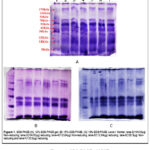 |
Figure 1: SDS-PAGE of MWE. |
Antithrombotic activity of MWE
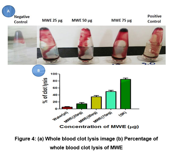
To determine the antithrombotic capabilities, in vitro plasma recalcification time, and in-vivo bleeding time were investigated. The study investigated the antithrombotic capabilities of MWE in human normal plasma. Figure 2 showed for both PPP, PRP, normal clotting time was 210 sec, and that the MWE demonstrated prolonged clotting time up to770 sec and exhibits anticoagulant activities.Each point represents the mean±SD of three independent experiments, P<0.02. Many research showed naturalproducts and their derivatives exhibits antithrombotic properties.41Additionally, Figure 3 represents MWE was verified by in vivo tail bleeding assay, the mice were injected with MWE solution in concentration gradient(0-100 µg) intravenously, which dramatically increased the bleeding time to 720 sec compare tocontrol. Prolonged bleeding time indicates anticoagulant property of MWE.Further, the role of MWE in the intrinsic/ extrinsic pathway of the blood coagulation cascade was confirmed by measuring APTT and PT. surprisingly MWE prolonged clotting time of APTT only without altering PT indicating it interferes intrinsic pathway of the blood coagulation cascade.Each point represents the mean±SD of three independent experiments, P<0.01From Marine creatures, herbal medicines, fermented food products like Japanese Natto and Korean Chungkook-Jang soy sauce, dung beetles, food-grade microorganisms,snake venoms, and earthworm secretions, several effective anticoagulant proteins/enzymes were identified by Mulenga A, et al. (2013).42 Figure 4. Represents, the amount of retraction is gradually inhibited by pre-incubating MWE with whole blood.90% of the blood clot was destroyed when MWE (0–100µg) was incubated against the positive control streptokinase (600U/ml). Each point represents the mean±SD of three independent experiments, P<0.01. Clot retraction assay confirms that MWE exhibits strong antithrombotic properties.All the above studies confirm antithrombotic properties of MWE. Several scientificreports showed milk and milk derived such as yogurt, cheese, etc. exhibit antithrombotic.43
 |
Figure 2: Plasma recalcification time of MWE |
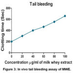 |
Figure 3: In vivo tail bleeding assay of MWE. |
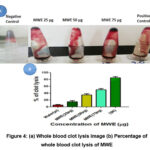 |
Figure 4: (a) Whole blood clot lysis image (b) Percentage of whole blood clot lysis of MWE |
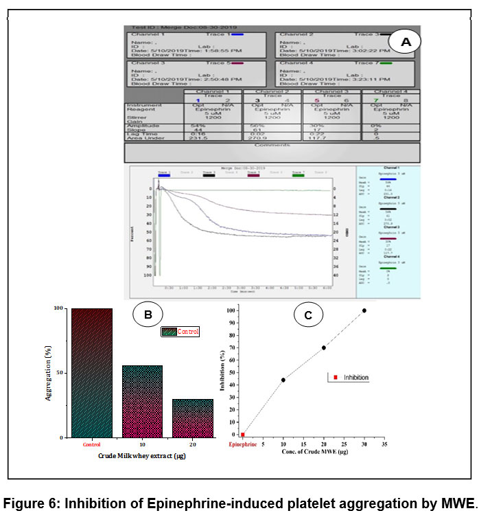
MWE inhibits platelet aggregation
The current study identified the effect of MWE on platelet aggregation was analysed using agonists such as ADP and epinephrine in PRP. Impressively, MWE at 30 µg concentration showed 74% of ADP induced inhibition (Figure 5) and 100% inhibition for epinephrine(Figure 6). Both agonists induced platelet aggregation inhibition at concentration of 0-30μg of MWE. Figure 5.represents(A)Traces of platelet aggregation, (B) dose dependent platelet aggregation%, (C) dose dependent platelet aggregation inhibition%.Figure 6. represents(A) Traces of platelet aggregation,(B) Dose dependent platelet aggregation%, (C) dose dependent platelet aggregation inhibition%. The values represent ± SD of three independent experiments.During the clotting mechanism in primary haemostasis, activation of platelets takes place by many agonists like ADP, thrombin, collagen, DAG, epinephrine, and platelet-activating factor (PAF). In thrombotic disorders, hyperactivation of platelets is commonly observed, whichis treated using Eptifibatide, a derivative from rattlesnake venom that inhibits glycoprotein IIb/IIIa receptor and so on Jung JL et al.(2013)44and Several platelet aggregation inhibitors have been identified from plants and animal sources.45-46Hence, aggregationresults show that MWE provestobea good therapeutic contender in the field of blood clotting /thrombotic disorders prevalence and treatments.
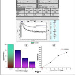 |
Figure 5: Inhibition of ADP-induced platelet aggregation by MWE |
 |
Figure 6: Inhibition of Epinephrine-induced platelet aggregation by MWE. |
Nontoxic property of MWE
MWE did not released haemoglobin by hemolysis of RBC showed in Figure 8. Packed RBC and PBS incubated with MWE concentration from 0-100 µg for 1 h at 37°C (Isotherm forced convection lab incubator, Inlab Equipments(Madras)Pvt. Ltd).Positive control: Water, negative control: PBS for percentage of hemolysis. The amount of hemoglobin released in the supernatant was measured at 540 nm(Thermo Nicolet iS50-Thermo Fisher Scientific, USA). Figure 7 showed MWE did not cause hemorrhage and edema was induced (data not revealed) in experimental mice up to the concentration of 100mg. Interestingly, MWE did not cause hemolysis as shown in fig. 8 when it was treated with RBC and did not cause hemorrhage and edema in experimental mice suggesting its non-toxic nature.The bioactive compounds found in MWE are thought to be interfering with the blood coagulation cascade based on the observations of all the aforementioned actions devoid of toxic properties.
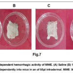 |
Figure 7: The dose-dependent hemorrhagic activity of MWE. (A) Saline (B) 125µg, |
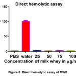 |
Figure 8: Direct hemolytic assay of MWE |
Conclusion
In summary, proximate analysis of MWE results found to be higher content of macronutrients carbohydrates and proteins, and micronutrients like vitamin A, potassium and calcium. In this study, an attempt was made to explore the antithrombotic and antiplatelet activities of MWE. Purified compounds of MWE proved to be good nutraceutical molecules and can be fortified with the meal to treat and manage cardiovascular and other life style disorders.Further purification and characterization of these bioactive compounds/ proteins appear to be interesting.
Acknowledgment
B.M and A.T thank the University Grant Commission, New Delhi.
Funding source
This research did not receive any specific grant from funding agencies in the public, commercial, or not-for-profit sectors.
Conflict of Interest
The authors declared no potential conflict of interest with respect to the authorship and publication.
Authors’ Contribution
Dr. Bhagyalakshmi M: -Research guide, guided me in all aspect of research.
Dr. Devaraja S: – Provided space in his research laboratory to carry out blood coagulation assays.
Jayanna K: – Assisted to carry out coagulation assays.
RameshaH: – Assisted to carry out proximate analysis.
Data Availability Statement
The manuscript incorporates all datasets produced or examined throughout this research study.
Ethics Approval Statement
This part is already mentioned above in the manuscript.
Reference
- Ahern DK, Gorkin L, Anderson JL, et al. Biobehavioral variables and mortality or cardiac arrest in the Cardiac Arrhythmia Pilot Study (Caps). The American Journal of Cardiology. 1990;66(1):59-62. doi:10.1016/0002-9149(90)90736-K.
CrossRef - Appels A, Mulder P. Fatigue and heart disease. The association between ‘vital exhaustion’ and past, present and future coronary heart disease. Journal of Psychosomatic Research. 1989;33(6):727-738. doi:10.1016/0022-3999(89)90088-3.
CrossRef - Carney RM, Rich MW, Freedland KE, et al. Major depressive disorder predicts cardiac events in patients with coronary artery disease.:Psychosomatic Medicine. 1988;50(6):627-633. doi:10.1097/00006842-198811000-00009.
CrossRef - Frasure-Smith N. Depression following myocardial infarction: impact on 6-month survival. JAMA. 1993;270(15):1819. doi:10.1001/jama.1993.03510150053029.
CrossRef - Affective disorders and survival after acute myocardial infarction*Results from the post-infarction late potential study. European Heart Journal. Published online September 1991. doi:10.1093/eurheartj/12.9.959.
CrossRef - Mortality among outpatients with anxiety disorders. AJP. 1986;143(4):508-510. doi:10.1176/ajp.143.4.508.
CrossRef - Haines AP, Imeson JD, Meade TW. Phobic anxiety and ischaemic heart disease. BMJ. 1987;295(6593):297-299. doi:10.1136/bmj.295.6593.297.
CrossRef - Park J, Lee B, Choi H, Kim W, Kim HJ, Cheong H. Antithrombosis activity of protocatechuic and shikimic acids from functional plant PinusdensifloraSieb. etZucc needles. J Nat Med. 2016;70(3):492-501. doi:10.1007/s11418-015-0956-y
CrossRef - The International Study Group. In-hospital mortality and clinical course of 20 891 patients with suspected acute myocardial infarction randomised between alteplase and streptokinase with or without heparin. The Lancet. 1990;336(8707):71-75. doi:10.1016/0140-6736(90)91590-7
CrossRef - Ohman EM, Harrington RA, Cannon CP, Agnelli G, Cairns JA, Kennedy JW. Intravenous thrombolysis in acute myocardial infarction. Chest. 2001;119(1):253S-277S. doi:10.1378/chest.119.1_suppl.253S
CrossRef - Hernández-Ledesma B, Del Mar Contreras M, Recio I. Antihypertensive peptides: Production, bioavailability and incorporation into foods. Advances in Colloid and Interface Science. 2011;165(1):23-35. doi:10.1016/j.cis.2010.11.001
CrossRef - Rafiq S, Huma N, Pasha I, Shahid M, Xiao H. Angiotensin‐converting enzyme‐inhibitory and antithrombotic activities of soluble peptide extracts from buffalo and cow milk Cheddar cheeses. Int J of Dairy Tech. 2017;70(3):380-388. doi:10.1111/1471-0307.12373
CrossRef - McGregor RA, Poppitt SD. Milk protein for improved metabolic health: a review of the evidence. NutrMetab. 2013;10(1):1
CrossRef - Massella E, Piva S, Giacometti F, Liuzzo G, Zambrini AV, Serraino A. Evaluation of bovine beta casein polymorphism in two dairy farms located in northern Italy. Ital J Food Safety. 2017;6(3). doi:10.4081/ijfs.2017.6904
CrossRef - Madureira AR, Pereira CI, Gomes AMP, Pintado ME, Xavier Malcata F. Bovine whey proteins – Overview on their main biological properties. Food Research International. 2007;40(10):1197-1211. doi:10.1016/j.foodres.2007.07.005
CrossRef - Aimutis WR. Bioactive properties of milk proteins with particular focus on anticariogenesis. The Journal of Nutrition. 2004;134(4):989S-995S. doi:10.1093/jn/134.4.989S
CrossRef - Fiat AM, Migliore-Samour D, Jollès P, Drouet L, Sollier CBD, Caen J. Biologically active peptides from milk proteins with emphasis on two examples concerning antithrombotic and immunomodulating activities. Journal of Dairy Science. 1993;76(1):301-310. doi:10.3168/jds.S0022-0302(93)77351-8
CrossRef - Hebert EM, Saavendra L, Ferranti P. Bioactive peptides derived from casein and whey proteins. Biotechnology of Lactic Acid Bacteria: Novel Applications.2010:233-249
CrossRef - Medeiros GKVV, Queiroga RCRE, Costa WKA, et al. Proteomic of goat milk whey and its bacteriostatic and antitumor potential. International Journal of Biological Macromolecules. 2018;113:116-123. doi:10.1016/j.ijbiomac.2018.01.200
CrossRef - Horwitz W, AOAC International, eds. Official Methods of Analysis of AOAC International. 18. ed., current through rev. 1, 2006. AOAC International; 2006.
- Khalid LP, Pulate PV, Wagay NA. Preliminary phytochemical analysis of miliusa tomentosa (roxb.) J. Sinclair by using various organi solvents. Euri Biomed Pharm SCI. 2017:4(5): 76-281. ISSN 2349-88A
- Sadeghzadeh S, Golgoli M, Masjoudi M, GhobadiNejad Z, Zargar M, Borghei SM. Removal of carbamazepine and diclofenac by laccase-based membrane bioreactor. Int J Environ Sci Technol. Published online February 18, 2024. doi:10.1007/s13762-023-05453-z
CrossRef - Laemmli UK. Cleavage of structural proteins during the assembly of the head of bacteriophage t4. Nature. 1970;227(5259):680-685. doi:10.1038/227680a0
CrossRef - Baburajeev CP, Mohan CD, Pandey V, et al. Synthesis of C C, C N coupled novel substituted dibutylbenzothiazepinone derivatives and evaluation of their thrombin inhibitory activity. Bioorganic Chemistry. 2019;87:142-154. doi:10.1016/j.bioorg.2019.03.004
CrossRef - Kengaiah J, Nandish SKM, Ramachandraiah C, Srinivasa C, Shivaiah A, Sannaningaiah D. In-vitro and in-vivo studies of Tamarind seed edible extract reveals anti-oxidative, anticoagulant, antiplatelet events. Asian J Pharm Pharmacol. 2019;5(6):1104-1116. doi:10.31024/ajpp.2019.5.6.5
CrossRef - Manjappa B, Gangaraju S, Girish KS, et al. Momordica charantia seed extract exhibits strong anticoagulant effect by specifically interfering in intrinsic pathway of blood coagulation and dissolves fibrin clot. Blood Coagulation & Fibrinolysis. 2015;26(2):191-199. doi:10.1097/MBC.0000000000000191
CrossRef - Denis C, Methia N, Frenette PS, et al. A mouse model of severe von Willebrand disease: Defects in haemostasis and thrombosis. Proc Natl AcadSci USA.1998;95(16):9524-9529. doi:10.1073/pnas.95.16.9524
CrossRef - Prasad S, Kashyap RS, Deopujari JY, Purohit HJ, Taori GM, Daginawala HF. Development of an in vitro model to study clot lysis activity of thrombolytic drugs. Thrombosis J. 2006;4(1):14. doi:10.1186/1477-9560-4-14
CrossRef - Ardlie NG, Han P. Enzymatic basis for platelet aggregation and release: the significance of the ‘platelet atmosphere’ and the relationship between platelet function and blood coagulation. Br J Haematol. 1974;26(3):331-356. doi:10.1111/j.1365-2141.1974.tb00477.x
CrossRef - Born GVR. Aggregation of blood platelets by adenosine diphosphate and its reversal. Nature. 1962;194(4832):927-929. doi:10.1038/194927b0
CrossRef - Kondo H, Kondo S, Ikezawa H, Murata R, Ohsaka A. Studies on the quantitative method for determination of hemorrhagic activity of habu snake venom. JJMSB. 1960;13(1-2):43-51. doi:10.7883/yoken1952.13.43
CrossRef - Pinheiro VDS, Souza MVGD, Oliveira GVD, et al. Physicochemical and protein profile of goat whey powder. Cienc Rural. 2024;54(1):e20220317. doi:10.1590/0103-8478cr20220317
CrossRef - Medeiros GKVV, Queiroga RCRE, Costa WKA, et al. Proteomic of goat milk whey and its bacteriostatic and antitumor potential. International Journal of Biological Macromolecules. 2018;113:116-123. doi:10.1016/j.ijbiomac.2018.01.200
CrossRef - Gomes JJL, Duarte AM, Batista ASM, et al. Physicochemical and sensory properties of fermented dairy beverages made with goat’s milk, cow’s milk and a mixture of the two milks. LWT – Food Science and Technology. 2013;54(1):18-24. doi:10.1016/j.lwt.2013.04.022
CrossRef - Palatnik DR, OstermannPorcel MV, González U, Zaritzky N, Campderrós ME. Recovery of caprine whey protein and its application in a food protein formulation. LWT – Food Science and Technology. 2015;63(1):331-338. doi:10.1016/j.lwt.2015.03.027
CrossRef - Kamizake NKK, Gonçalves MM, Zaia CTBV, Zaia DAM. Determination of total proteins in cow milk powder samples: a comparative study between the Kjeldahl method and spectrophotometric methods. Journal of Food Composition and Analysis. 2003;16(4):507-516. doi:10.1016/S0889-1575(03)00004-8
CrossRef - Jung TH, Yun SS, Lee WJ, et al. Hydrolysis by alcalase improves hypoallergenic properties of goat milk protein. Korean Journal for Food Science of Animal Resources. 2016;36(4):516-522. doi:10.5851/kosfa.2016.36.4.516
CrossRef - Caillat H, Bouvier F, Martin P. Major proteins of the goat milk fat globule membrane. J Dairy Sci. 2010;3(93):868-876
CrossRef - 李永祯, 黄大通, 邢世其, 王雪松. 合成孔径雷达干扰技术研究综述. ldxb. 2020;9(5):753-764. doi:10.12000/JR20087
CrossRef - Somaye HN, Firoozeh HE, Golaleh A, et al.Nutrition and Cardiometabolic Risk Factors: Findings from 20 Years of the Tehran Lipid and Glucose Study; 2018.
CrossRef - Fiat AM, Migliore-Samour D, Jollès P, Drouet L, Sollier CBD, Caen J. Biologically active peptides from milk proteins with emphasis on two examples concerning antithrombotic and immunomodulating activities. Journal of Dairy Science. 1993;76(1):301-310. doi:10.3168/jds.S0022-0302(93)77351-8
CrossRef - Mulenga A, Kim T, Ibelli AMG. amblyommaamericanum tick saliva serine protease inhibitor 6 is a cross‐class inhibitor of serine proteases and papain‐like cysteine proteases that delays plasma clotting and inhibits platelet aggregation. Insect Molecular Biology. 2013;22(3):306-319. doi:10.1111/imb.12024
CrossRef - Al-Anazi MS, El-Zahar KM, Rabie NAH. Nutritional and therapeutic properties of fermented camel milk fortified with red chenopodium quinoa flour on hypercholesterolemia rats. Molecules. 2022;27(22):7695. doi:10.3390/molecules27227695
CrossRef - Alfonsi E, Méheust E, Fuchs S, et al. The use of DNA barcoding to monitor the marine mammal biodiversity along the French Atlantic coast. ZK. 2013;365:5-24. doi:10.3897/zookeys.365.5873
CrossRef - Bhagyalakshmi M, Devaraja S. Momordica charantia (Bitter melon): Potent antiviral efficacy and significant benefits against herpes virus. In: Viral, Parasitic, Bacterial, and Fungal Infections. Elsevier; 2023:209-220. doi:10.1016/B978-0-323-85730-7.00017-5
CrossRef - Denis C, Methia N, Frenette PS, et al. A mouse model of severe von Willebrand disease: Defects in hemostasis and thrombosis. Proc Natl AcadSci USA. 1998;95(16):9524-9529. doi:10.1073/pnas.95.16.9524
CrossRef
Abbreviations
ADP : Adenosine diphosphate
APTT : Activated partial thromboplastin time
AOAC : Association of Official Agricultural Chemists
BSA : Bovine serum albumin
DAG : Diacylglycerol
FC : Folin-Ciocalteu’s
g : Gram
h : Hour
ICP-OES : Inductively coupled plasma optical emission spectrometry
kDa : Kilo Dalton
L : Litre
MWE : Milk whey extract
mg : Milligram
ml : Millilitre
µg : Microgram
min : Minute
mM : Millimolar
mm : Millimetre
PAF : Platelet-activating factor
PBS : Phosphate buffered saline
PPP : Platelet-poor plasma
PRP : Platelet rich plasma
PRT : Plasma recalcification Time
PT : Prothrombin time
RBC : Red blood cells
rpm : Revolution per minute
SDS : Sodium dodecyl sulfate
Sec : Second
wt : Weight

This work is licensed under a Creative Commons Attribution 4.0 International License.


