
Introduction
Japanese apricot is a deciduous small tree of the Rosaceae family that originated from the southwest of China. It has a long history of cultivation in China and has been introduced to Taiwan, Japan, Korea, Vietnam, and other countries1 because of its high ornamental value. Japanese apricot is rich in nutrients and contains a variety of essential minerals and organic acids2,3 that have beneficial effects on health. The consumption of Japanese apricots as a health food is widespread in Asian countries such as China and Japan. However, fresh and unprocessed Japanese apricots are randomly consumed because of their severe sourness and astringency. 4
The Japanese apricot has been used in traditional medicine for more than 3000 years and was used to treat various symptoms. Plant parts, such as branches, leaves, flowers, seeds, and roots, have been used in traditional medicine in China, but most pharmacological studies have been performed with fruit extracts. Fructus mume, produced from the unripe fruit of Prunus mume, is one of the most famous and ancient medicinal herbs and health foods used in China.5
According to traditional Chinese herbal medicine records, Fructus mume has been used to treat various diseases, such as chronic diarrhea, cough, expectoration, ulceration, gastrointestinal diseases, and nocturnal asthma symptoms. 6-13 Moreover, it is also used as one component in many formulas, and modern studies have reported that Fructus mume exerts a therapeutic effect on various diseases owing to its anti-inflammatory, antioxidant, antibacterial, and radical scavenging activities. 12,14 The components in Fructus mume are complex and diverse. Like the fresh fruit of Prunus mume, it contains various nutritional and functional substances, such as organic acids and minerals. But there is no report on the consistency of organic acids and mineral elements between Fructus mume and fresh fruits of Prunus mume. To the best of our knowledge, ultra-performance liquid chromatography (UPLC), inductively coupled plasma atomic emission spectrometer (ICP-OES), and inductively coupled plasma source mass spectrometer (ICP-MS) were used to determine the types and contents of organic acids and mineral elements in Fructus mume.
The microbiota plays a major role in health and disease, It participates in various metabolic functions such as intestinal homeostasis, and development. and that exists in the intestinal flora is associated with metabolic reactions and immunomodulatory.15-18 It is reported that dietary changes show significant impacts on the microbiota.19 Previous studies have reported that Prunus mume fruit juice concentrate could affect the gut microbiota in mice.20 However, the effects of Fructus mume on the gut microbiota have not been investigated. Therefore, this study aimed to investigate the effects of Fructus mume on the intestinal flora by 16s rRNA sequencing. The liver and kidney tissues were used to discuss the effects of Fructus mume on the liver and kidney of mice in detail.
Materials and Methods
Experimental materials
The Fructus mume juice used in this study was provided by the National Field of Waxberry and Prunus mume at the Nanjing Agricultural University, Nanjing, Jiangsu Province.
Chemicals
The reagents used included chromatography-grade standards for oxalic, tartaric, malic, ascorbic, acetic, citric, maleic, and fumaric acid (organic acid); mineral element standards for potassium (K), phosphorus (P), calcium (Ca), magnesium (Mg), sodium (Na), selenium (Se), and germanium (Ge); disodium hydrogen phosphate; phosphoric acid; and a 4% polyformaldehyde fixing solution.
UPLC quantitative analysis of the organic acid
The organic acid content was accurately determined by UPLC, which consisted of a Waters ACQUITY UPLC HSS T3 column (2.1 mm×100 mm, 1.8μm). The mobile phase was 50 mM sodium dihydrogen phosphate (pH 2.4). The flow rate was set at 0.25 mL/min, the column temperature was maintained at 30 ℃, the injection volume was 2 μL, the PAD was used as the detector, and the detection wavelength was 214 nm.
ICP-OES and ICP-MS quantitative analysis of the mineral element
The mineral element content was accurately measured by ICP-OES and ICP-MS.
Preparation and determination of mineral element standard solutions: Standard solutions of Ca, K, and Na with a concentration of 1000 mg/L were diluted to 10 mg/L, 5 mg/L, 2.5 mg/L, 1.25 mg/L, and 0.625 mg/L, respectively. The standard solutions of Mg, P, Se, and Ge with a concentration of 1000 mg/L were diluted to 10 mg/L, 5 mg/L, 2 mg/L, 1 mg/L, and 0.5 mg/L. The blank sample was made, and all the samples were measured on the machine.
Feeding and gavage of mice
The SPF male healthy mice (4 weeks) were used in this study with a weight between 18-20 g. All experimental mice were obtained from the Experimental Animal Center of Nanjing Agricultural University and were raised in the barrier environment animal room of the Experimental Animal Center of Nanjing Agricultural University. This study was approved by Animal Ethics Committee of Nanjing Agricultural University Permit (NJAU. No20221031206) and performed in accordance with the guidelines of the Science and Technology Agency of Jiangsu Province and Nanjing Agricultural University.
A total of 12 mice were randomly divided into two groups (n=6): the CK group with treatment of distilled water and the other group with Fructus mume juice. They were fed and drank adaptively for one week and then given gavage continuously six times a week for six weeks. The mental state, activity, and foraging amount of the mice were observed after gavage, and the weight of the mice was measured once a week.
The fresh manure of mice was collected after 12 h.
High-throughput sequencing of intestinal flora for mouse
Stool samples from each group (n=6/group) were collected and stored at -80 ℃ for further analysis. The samples were completed by Nanjing Zhongke Shikang Biotechnology Co., Ltd. (Nanjing, China). The total DNA of the specimen was extracted from a DNA extraction kit. A database was created for qualified DNA samples. PCR amplification was performed using Pfu high-fidelity DNA polymerase from the Total Gold Company, using microbial ribosomal RNA or specific gene fragments and other target sequences that could reflect the composition and diversity of the flora as the target. The corresponding primers were designed, and the quality of the products was assessed by a Microplate reader (BioTek, FLx800). The library was sequenced using a two-terminal sequencing strategy on the MiSeq platform, and quality control was performed using QIIME2, USEARCH, and VSearch. The operational taxonomic units (OTUs) were identified by QIIME2. Community bar-pot, alpha and Beta diversity analyses, heat mapping, and species identification were performed using QIIME2 and R software packages (http://www.R-project.org).
Determining the response of liver and kidney in mice
Mouse eyeball blood was collected, incubated at 4 °C for 30 min, and centrifuged at 3000 r/min for 20 min, and the supernatant was used for testing the ALT, AST, BUN, and CR using test kits.
After blood collection, the kidney and liver tissues were collected for pathological observation; the mice were sacrificed by neck removal, and liver and kidney tissues were collected for experiments. The collected tissues were weighed and then fixed in a 4% polyformaldehyde solution and dyed. The formula for calculating the organ coefficient was as follows: The organ coefficient = (organ wet mass/mouse body mass) *100%.
Statistical analysis
The statistics were analyzed using Excel and SPSS (version 26.0). All data were expressed as mean ± standard deviation. Comparisons were based on t-tests of the independent samples. A p-value<0.05 was considered statistically significant.
Results
The functional components in Fructus mume
The composition and content of organic acids in Fructus mume
The main nutrition in the fruit of Japanese apricot is organic acid, and there are eight types of organic acid in it. In this study, eight kinds of organic acids were determined in Fructus mume, similar to those in the fresh fruit of Japanese apricots. The total organic acid content in Fructus mume was measured at 31.73 g/L. The main kind of organic acid in Fructus mume was citric acid, and the content was 25.35±0.049 g/L, accounting for 79.89% of the total organic acid. The malic acid was the secondary major with a 2.68±0.01g/L content. The succinic acid content was recorded at 0.019±0.001g/L, the lowest among the eight kinds of organic acid (Figure 1).
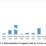 |
Figure 1: Determination of organic acids in Fructus mume. |
Mineral elements in Fructus mume
The seven types of mineral element and their content ranges are listed in Figure 2. The result showed that the content of the seven mineral elements in Fructus mume followed the order from highest to lowest K>Ca>P>Na>Mg>Se>Ge. Similar to Prunus mume fruit, which is rich in potassium, phosphorus, calcium, magnesium, and selenium.21 As is seen, potassium content, recorded at 4566.03±130.76 mg/L, was the highest in Fructus mume, accounting for 79.9% of all the determined mineral element content.
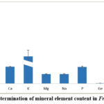 |
Figure 2: Determination of mineral element content in Fructus mume. |
The effect of Fructus mume on the body weight of mice
Mice in the control and Fructus mume groups showed normal behavior and activity levels. There were no significant differences in mice body weights before the experiment, and the final body weight of mice in the Fructus mume group was higher than that of the control group (Table 1). However, no significant differences were observed between the two treatments.
Table 1: Effects of Fructus mume on body weight of mice
| Group | 0week | 1 week | 2 weeks | 3 weeks | 4 weeks | 5 weeks | 6 weeks | Weight gain |
| Control | 19.2±0.64 | 19.6±0.71 | 19.9±1.26 | 19.9±1.32 | 19.6±1.48 | 20.0±1.54 | 20.9±0.82 | 1.7±1.10 |
| Fructus mume | 19.3±0.62 | 19.2±0.82 | 20.2±0.83 | 20.4±0.63 | 20.7±1.08 | 21.5±1.22 | 21.6±1.54 | 2.3±2.00 |
Note: * indicates significant differences in the same column.
Effects of Fructus mume on intestinal flora of mice
Information on sequencing and species composition
To investigate the effects of Fructus mume on the intestinal flora of mice, 16S rRNA high-throughput sequencing was used in this study. 3297342 original sequences were obtained. There were 2899159 effective sequences after noise removal and 1681057 high-quality sequences after chimera removal, with an average of 67501 sequences per sample. The length distribution of all high-quality sequences was analyzed, and the results showed that the length was mainly concentrated in the 404-432 bp (Figure 3).
 |
Figure 3: Distribution diagram of sequence length. |
As shown in the OTUs Venn diagram obtained from the cluster of the two groups of samples, the OUTs number of the Fructus mume group was 6822, which was higher than that of the control group (6306). A total of 354 species were found in both groups (Figure 4). As shown in the map of the specific composition of microbial communities at each taxonomic level, the number of taxa in the two treatments was calculated at each classification level. The results showed that the class, order, family, genus, and species levels of the Fructus mume group were higher than those of the control group.
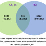 |
Figure 4: Venn diagram illustrating the overlap of OUTs for intestinal flora in mice. Blue represents the Fructus mume group (FM) and green represents the control group (CK_m). |
Analysis of flora composition and species diversity at phyla and genus levels
According to the results of species annotation, the top 10 species in the relative abundance of each group at the phylum and genus levels were selected (Figure 5) in this study. At the phylum level, the predominant phyla in the mouse gut were Bacteroidetes and Firmicutes, accounting for an average proportion of > 87%. Other dominant phyla were Actinobacteria, TM7, Tenericutes, Proteobacteria, Verrucomicrobia, Cyanobacteria, Deferribacteres, and Acidobacteria. The abundance ratio of Firmicutes and Bacteroidetes of the Fructus mumue group was higher than that of the control group, which is the same as the changing trend of the body mass of the mice. The results showed that Fructus mume modulated the Firmicutes to Bacteroides ratio, optimized intestinal microbiota composition in mice, enhanced the proliferation of beneficial bacteria, improved intestinal flora function, and promoted body weight gain in mice.
The dominant bacteria genera at the genus level are Lactobacillus, Bacteroides, Oscillospira, Ruminococcus, Adlercreutiza, Odoribacter. Allobaculum, Parabacteroides, Alistipes, and Enterococcus. Compared with the control group, the relative abundance of Lactobacillus, which played a role in preserving the microecological equilibrium with gastrointestinal traces, was significantly increased. The relative abundances of Oscillospira, Adlercreutzia, Odoribacter, Allobaculum, Alistipes, and Enterococcus were enhanced in the Fructus mume group. At the same time, those of Ruminococcus and Parabacteroides were lower in the Fructus mume group.
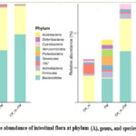 |
Figure 5: Relative abundance of intestinal flora at phylum(A), genus, and species (B) level in mice. |
Analysis of Alpha diversity of intestinal flora in mice
Shannon and Simpson indices and richness of Chao1 and Observed_species of the gut microbiota characterized alpha diversity.
Goods_coverage indices of the two groups were higher than 0.98, suggesting that the sequencing depth was sufficient to cover all sample species. To evaluate the diversity and richness of intestinal flora, we analyzed alpha diversity through Chao1, Observed Species, Shannon, and Simpson Pielou indices (Table 2), and the boxplot of the alpha diversity index is shown in Figure 6. The richness of the mice in the Fructus mume group was lower than that in the control group. There were significant differences in the Pielou indices and no significant differences in the Chao 1, Observed_species, Shannon, and Simpson indices between the two groups.
Table 2: Richness and microbial diversity index of flora from mice.
| Group | Chao1 | Simpson | Shannon | Pielou | Observed_species | Goods_coverage | ||
| Control | 2277.51±434.33 | 0.97±0.00 | 7.34±0.40 | 0.67±0.01 | 1970.4±482.05 | 0.99±0.00* | ||
| Fructus mume | 2692.58±307.68 | 0.98±0.01 | 7.76±0.25 | 0.7±0.02* | 2130.93±258.90 | 0.98±0.00 | ||
Note: * indicates significant differences in the same column.
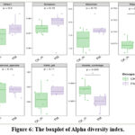 |
Figure 6: The boxplot of Alpha diversity index. |
Analysis of Beta diversity of intestinal flora in mice
Beta diversity, also known as habitat diversity, refers to the variation in species composition between different communities along environmental gradients or the rate of species turnover along these gradients. Multi-dimensional microbial data can be reduced through unconstrained sorting techniques, such as principal coordinate analysis (PCoA) and non-metric multidimensional scaling (NMDS), to visualize the main trends of data changes by displaying the distribution of samples along continuous sorting axes. Clustering analysis can also be used to identify discrete subsets of objects in an environment and classify the data.
NMDS was performed to determine the beta diversity of the different groups. The greater the distance between various groups, the greater the difference in diversity.
The Fructus mume group was separated from the control group, suggesting significant differences between the two groups in the species structure composition of the intestinal flora. In conclusion, the beta diversity of the Fructus mume group was higher than that of the control group, and Fructus mume enhanced the beta diversity of the intestinal flora of mice (Figure 7).
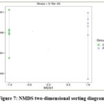 |
Figure 7: NMDS two-dimensional sorting diagram. |
Effect of Fructus mume on the liver and kidney of mice
Coefficient of liver and kidney
The coefficients of the liver and kidney were tested in this study to evaluate the effect of the Fructus mume. As is seen in Table 3, the liver coefficient of the Fructus mume group was 5.50±0.16%, higher than that of the control group; the kidney coefficient of the Fructus mume group was 1.57±0.07 %, and it was higher, too. However, the two treatments had no significant differences in liver and kidney coefficients.
Table 3: Effect of Fructus mume juice on liver and kidney coefficient of mice.
| Group | Liver coefficient(%) | Kidney coefficient(%) |
| Control | 4.98±0.47 | 1.44±0.12 |
| Fructus mume | 5.50±0.16 | 1.57±0.07 |
Note: * indicates significant differences in the same column.
Pathological observation of liver and kidney
The liver and kidney tissues of mice treated with distilled water (Control group) and Fructus mume juice (Fructus mume group)were examined in this study (Figures 8 and 9). The liver and kidney structures of mice in the control and Fructus mume groups were normal. In this study, we found no obvious damage.
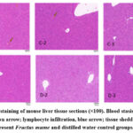 |
Figure 8: HE staining of mouse liver tissue sections (×100). Blood stasis, black arrow; bleeding, brown arrow; lymphocyte infiltration, blue arrow; tissue shedding, red arrow. C and D represent Fructus mume and distilled water control groups, respectively. |
The renal tissues of mice in the two treatments showed no apparent damage, and their structures were typical. Some differences were observed in the mouse cortex. However, a small amount of tissue bleeding in some areas of the cortex and lymphocyte infiltration in some areas were observed in the Fructus mume group. There was a small area of lymphocyte infiltration in the medullary substance of mice in the Fructus mume group.
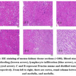 |
Figure 9: HE staining of mouse kidney tissue sections (×100). Blood stasis (black arrow), bleeding (brown arrow), lymphocyte infiltration (blue arrow), and tissue shedding (red arrow). |
Determination of serum biochemical factors
Glutamic-pyruvic transaminase(AST) and glutamic oxaloacetic transaminase (ALT) are commonly used to assess liver damage. Both the ALT and AST increased in the case of liver cell membrane permeability or liver cell necrosis, and blood urea nitrogen (BUN) and creatinine(CR) were used to evaluate the extent of kidney damage. In this study, we determined ALT and AST levels to evaluate liver damage and BUN and CR levels in the kidneys.
Based on the liver function indices, the ALT and AST levels of the mice in the Fructus mume group were lower than those in the control group. However, there were no significant differences between the ALT levels of the two groups, but there was a significant difference in the AST levels between the two groups. Compared with the control group, the levels of BUN and CR in the Fructus mume group were lower, and the CR content of that group was significantly lower. After the experiment, the mice were in good physical and mental condition.
Table 4: Effect of Fructus mume juice on serum biochemical factors of mice
| (Group) | ALT(U/L) | AST(U/L) | BUN(mg/dl) | CR(μmol/L) |
| Control | 49.49±7.13 | 118.78±14.44 | 13.61±2.23 | 22.72±1.27 |
| Fructus mume | 47.64±19.87 | 98.18±12.56* | 11.74±1.28* | 18.42±1.34 |
Note: * indicates significant difference in the same column.
Discussion
Advances in food and nutrition have shifted consumer preferences towards nutraceutical and functional foods.22 Fructus mume, processed from the unripe fruit of Japanese apricot by drying at low temperatures,8,23 is a natural food with nutritive and pharmaceutical value and has been used as a medicinal and functional food in Asian countries for more than 3000 years to treat a series of diseases 6-13. The functional components in the fruit of Prunus mume include organic acids, mineral elements, fiber, polysaccharides, flavonoids, and polyphenol compounds.3,24 In our study, the concentrations of eight kinds of organic acids in the fruit of Fructus mume were evaluated quantitatively by UPLC to determine the differences between the processed and fresh fruit. The results showed that the content of organic acids in Fructus mume was higher than that in the fruit of Prunus mume. Similar to Prunus mume fruit, citric acid, the Q-marker of Fructus mume in the Chinese Pharmacopoeia,9,23 was the primary type of organic acid in Fructus mume, accounting for 79.89% of the total content determined (Figure 1). It also has antioxidant, antibacterial, antithrombotic, and anti-inflammatory effects.23 Malic acid, which accounted for 8.44%, was the secondary main kind of organic acid and higher than that of Prunus mume fruit.
Minerals are required as essential nutrients by humans to carry out the functions required for health.25-26 The contents of mineral elements in Fructus mume were higher than those in Prunus mume fruit. The K content was the highest among the seven mineral elements determined in this study. Potassium is involved in regulating water, electrolyte, and acid-base balance in the body.27-29 This study found that K was the main mineral element, accounting for 70% of the total content.
It has been confirmed that ten phylogenetic bacterial groups exist in the intestinal microbiota.30 The composition and diversity of gut microbiota are related to health and disease.31 The gut microbiota performs essential metabolic functions in conjunction with the host’s defense and immune systems to protect against pathogen infiltration and colonization.19,32 The imbalance of it can lead to the occurrence of many diseases, such as obesity,33-34 hypertension,35 and cardiovascular disease.32 Therefore, 16S rRNA sequencing was used to analyze the composition of the intestinal flora of the mice in this study. The results showed that the intestinal flora composition of Fructus mume was different from that of the control group. Firmicutes and Bacteroidetes are the main bacterial phyla, and the F/B ratio plays a crucial role in maintaining normal intestinal homeostasis.18,36
The genus Bacteroides is enriched in the colon of chronic kidney disease patients with CKD.20,37 Our study found that the abundance of Bacteroides of Fructus mume group was lower than that in the control group, and the BUN and CR contents of the former group were also significantly lower than those of the control group (Table 4). In conclusion, we can deduce that Fructus mume is beneficial in protecting the kidneys from damage.
Lactobacillus is known to have probiotic effects in animals,38-39 the abundance of which could slow the progression of kidney disease by improving the intestinal environment 20,40. We found that the abundance of this species was greater in the Fructus mume group, which implies that Fructus mume might promote the multiplication of Lactobacillus species. Oscillospira has a positive effect on human health, low fat, leanness, and inflammatory disease of the liver,41-42 and in this study, we find that the relative abundance of Oscillospira is significantly higher compared with control.
Conclusion
In summary, Fructus mume intake could increase the body weight of mice and enrich the abundance of intestinal flora at the phylum and genus levels. It was beneficial to protect the liver and kidney from damage by reducing the serum biochemical factors. This study quantitatively analyzed the organic acids and minerals of Fructus mume, which are the main functional components in Japanese apricot fruit, using UPLC, ICP-OES, and ICP-MS. The organic acid in Fructus mume was higher than in Prunus mume fruit. Similar to Prunus mume, the primary mineral element in Fructus mume is K . These findings provide theoretical support for further exploration of the medicinal value of Fructus mume.
Acknowledgement
The authors would like to thank the Experimental Animal Center of Nanjing Agricultural University for providing the experimental mice.
Funding Sources
This work was financial supported by the Chongqing Jiangji Winery Co., Ltd, the Fundamental Research Funds for the Central Universities, and the Priority Academic Program Development of Jiangsu Higher Education Institutions (PAPD).
Conflict of Interest
The authors declare no conflict of interest.
Data Availability Statement
The manuscript incorporates all datasets produced throughout this research study.
Ethics Statement
This study was approved by Animal Ethics Committee of Nanjing Agricultural University Permit and performed in accordance with the guidelines of the Science and Technology Agency of Jiangsu Province and Nanjing Agricultural University.
Informed Consent Stetement
This study did not involve human participants, and therefore, informed consent was not required.
Permission to reproduce material from other sources
This study did not contains any materials from other source.
Clinical Trial Registration
This research does not involve any clinical trials.
Author Contributions
- Jing Shao: Writing original draft, Investigation, Formal analysis, Data curation.
- Shasha Wang: Writing original draft, Investigation, Formal analysis, Data curation.
- Jiangfeng Song: Formal analysis and reviewing multiple versions of the manuscript.
- Xueming Zhang: Formal analysis, Writing-Review& Editing.
- Faisal Hayat: Investigation, Data curation.
- Hengguang Li: Providing funding support and suggestions.
- Wenjin Li: Providing funding support and suggestions.
- Zhihong Gao: Formal analysis, Conceptualization, Funding acquisition.
References
- Bailly C, Anticancer properties of Prunus mume extracts (Chinese plum, Japanese apricot), J Ethnopharmacol., 2020, 246, 112215.
CrossRef - Chu MY, China fruit records–mei. China Forestry Press, 1999, Beijing.
- Gao ZH, Shao J, Sun HL, et al. Evaluation of different kinds of organic acids and their antibacterial activity in Japanese Apricot fruits. J. Agric. Res., 2012, 7(35), 4911-4918.
CrossRef - Tian TT, Cao H, Farag MA, et al. Current and potential trends in the bioactive properties and health benefits of Prunus mume Et Zucc: a comprehensive review for value maximization. Cri. Rev in Food Sci. and Nutr., 2023, 63(24), 7091-7107.
CrossRef - Gong XP, Tang Y, Song YY, et al. Comprehensive review of phytochemical constituents, pharmacological properties, and clinical applications of Prunus mume, Pharmacol., 2021, 12, 679378.
CrossRef - Tang LL, Xu YQ, Yang Y, et al. Identification of the role of Mume Fructus in treating nocturnal asthma based on network pharmacology and in vitro investigation, World J. Tradit. Chin. Med., 2024, 10, 4103.
CrossRef - Zhao MJ, Zhao Q, Guan ZW, et al. Effect of Panax ginseng and Fructus Mume on intestinal barrier and gut microbiota in rats with diarrhea, Med. Food., 2023, 26(3), 165-175.
CrossRef - Xu ZY, Zhang XL, Wang WY, et al. Fructus Mume (Wu Mei) attenuates acetic acid-Induced ulcerative colitis by regulating inflammatory cytokine, reactive oxygen species, and neuropeptide levels in model rats, Med. Food., 2022, 25, 389–401.
CrossRef - Liu ZH, Peng Y, Ma P, et al. An integrated strategy for anti-inflammatory quality markers screening of traditional Chinese herbal medicine Mume Fructus based on phytochemical analysis and anti-colitis activity., 2022, 99, 154002.
CrossRef - Xu ZM, Zhang XL, Lu RM, et al. Mechanism of Fructus Mume pills underlying their protective effects in rats with acetic acid-inducedulcerative colitis via the regulation of inflammatory cytokines and the VEGF-PI3K/Akt-eNOS signaling pathway, Evid-Based Complement. Med., 2022, 4621131.
CrossRef - Xiang J, Liu XD, Zhong S, et al. Fructus mume protects against cigarette smoke induced chronic cough guinea pig. Med. Food., 2020, 23, 191–197.
CrossRef - Kim MS, Bang JH, Lee J, et al. Fructus mume ethanol extract prevents inflammation and normalizes the septohippocampal cholinergic system in a rat model of chronic cerebral hypoperfusion. Med. Food., 2016, 19(2), 196–204.
CrossRef - Xing H, Zhang LR, Ma JS, et al. Fructus mume extracts alleviate diarrhea in breast cancer patients receiving the combination therapy of lapatinib and capecitabine. Pharmacol., 2018, 9, 516.
CrossRef - Debnath T, Bak JP, Samad NB, et al. Antioxidant activity of mume Fructus J. Food Biochem., 2012, 36(2), 224-232.
CrossRef - O’Hara, A.M., and Shanahan, F. The gut flora as a forgotten organ. EMBO Rep., 2006, 7, 688–693
CrossRef - Stojanov S, Berlec A, Štrukelj B. The influence of probiotics on the firmicutes/bacteroidetes ratio in the treatment of obesity and inflammatory bowel disease. Microorganisms, 2020, 8(11), 1715.
CrossRef - Thursby E, Juge N. Introduction to the human gut microbiota. J., 2017 , 474, 1823–1836.
CrossRef - Montalto M, D’Onofrio F, Gallo A,et al. Intestinal microbiota and its functions. Dig. Liver. Dis., 2009, 3, 30–34.
CrossRef - Clemente JC, Ursell LK, Parfrey L, et al. The impact of the gut microbiota on human health: an integrative view. Cell, 2012, 148(6), 1258-1270.
CrossRef - Huang Y, Wu CX, Guo L, et al. Effects of polysaccharides-riched Prunus mume fruit juice concentrate on uric acid excretion and gut microbiota in mice with adenine-induced chronic kidney disease. Curr. Res. Food Sci., 2022, 5, 2135-2145.
CrossRef - Ali S, Masud T, Abbasi KS, et al.Apricot: nutritional potentials and health benefits-a review. Food Sci. Tech., 2015, 16: 175-189.
- Fatima T, Bashir O, Gani G, Bhat TH. Nutritional and health benefits of apricots. Int. J. Unani. Integ. Med., 2018, 2(2), 5-9.
CrossRef - Gao L, Zhang H, Wang H, et al. Effects of different varieties on physicochemical properties, browning characteristics, and quality attributes of Mume fructus (Wumei). Foods, 2024, 13(9), 1377.
CrossRef - Wang YK, Li AT, Huang X, et al. Chemical profiling and antioxidant activity of Japanese apricot flowers with green sepals: Insights into medicinal potential and harvest optimization. Ind Crops and Prod., 2024, 212, 118324
CrossRef - Godswill AG, Somtochukwu I V, Ikechukwu AO, et al. Health benefits of micronutrients (vitamins and minerals) and their associated deficiency diseases: A systematic review. J. Food Sci., 2020, 3(1), 1-32.
CrossRef - Prashanth L, Kattapagari KK, Chitturi RT, et al. A review on role of essential trace elements in health and disease. Ntr. Univ. Health Sci., 2015, 4(2), 75-85.
CrossRef - Czech A, Zarycka E, Yanovych D, et al. Mineral content of the pulp and peel of various citrus fruit cultivars. Bio. Trace Elem. Res., 2020, 193, 555-563.
CrossRef - Stone MS, Martyn L, Weaver CM. Potassium intake, bioavailability, hypertension, and glucose control. Nutrients, 2016, 8(7): 444.
CrossRef - Pohl HR, Wheeler JS, Murray HE. Sodium and potassium in health and disease. In: Sigel A, Sigel H, Sigel RKO (eds) Interrelations between essential metal ions and human diseases, Springer Netherlands, Dordrecht, 2013, pp 29–47
CrossRef - Tamura M, Ohnishi Y, Kotani T. Effects of new dietary fiber from Japanese Apricot (Prunus mume et Zucc.) on gut function and intestinal microflora in adult mice. Int. J. Mol. Sci., 2011, 12(4), 2088-2099.
CrossRef - Larsen OF, Claassen E. The mechanistic link between health and gut microbiota diversity. Sci. Rep., 2018, 8(1), 2183.
CrossRef - Witkowski M, Weeks TL, Hazen SL. Gut microbiota and cardiovascular disease. Circ. Res., 2020, 127(4), 553-570.
CrossRef - Mitev K, Taleski V. Association between the gut microbiota and obesity. Open access Maced. J. Med. Sci., 2019, 7(12), 2050-2056.
CrossRef - Amabebe E, Robert FO, Agbalalah T, et al. Microbial dysbiosis-induced obesity: role of gut microbiota in homoeostasis of energy metabolism. J. Nutr., 2020, 123(10), 1127- 1137.
CrossRef - Li J, Zhao FQ, Wang YD, et al. Gut microbiota dysbiosis contributes to the development of hypertension. Microbiome, 2017, 5(4), 1-19.
CrossRef - Sutoyo DA, Atmaka DR., Sidabutar LMGB. Dietary factors affecting Firmicutes and Bacteroidetes ratio in solving obesity problem: a literature review. Media Gizi Indones., 2020, 15(2), 94-109.
CrossRef - Eraly SA, Vallon V, Rieg T, et al. Multiple organic anion transporters contribute to net renal excretion of uric acid. Physiol. Genom., 2008, 33, 180–192
CrossRef - Karunasena E, Kurkure PC, Lackey RD, et al. Effects of the probiotic Lactobacillus animalis in murine Mycobacterium avium subspecies paratuberculosis infection. BMC Microbiol.,2013, 13, 8.
CrossRef - Mayer MJ, D’Amato A, Colquhoun IJ, et al. Identification of genes required for glucan exopolysaccharide production in Lactobacillus johnsonii suggests a novel biosynthesis mechanism. Appl. Environ. Microbiol., 2020.86(8), e02808-19.
CrossRef - Yoshifuji A, Wakino S, Irie J, et al. Gut Lactobacillus protects against the progression of renal damage by modulating the gut environment in rats. Nephrol. Dial. Transplant., 2016, 31, 401–412
CrossRef - Konikoff T, Gophna U. Oscillospira: a central, enigmatic component of the human gut microbiota. Trends Microbiol., 2016, 24(7), 523-524.
CrossRef - Yang J, Li Y, Wen Z, et al. Oscillospira-a candidate for the next-generation probiotics. Gut microbes, 2021, 13(1), 1987783.
CrossRef

This work is licensed under a Creative Commons Attribution 4.0 International License.


