Introduction
Peanut is one of the most important grain legumes extensively cultivated globally, primarily used in confectionaries and for edible oil production. During the peanut blanching process, the industry removes substantial amounts of potentially polluting peanut skin, which is a primary residue and by-product1 having low commercial value, and limited industrial applications, mainly being used as livestock feed.2 The skins are pink-red in colour and are nontoxic layer that covers peanuts.3 Nevertheless, peanut skin is an excellent raw material containing phenolic compounds, particularly proanthocyanidins.4,5 Other major classes of natural phenolics such as stilbenes, phenolic acids and flavonoids have also been found in peanut skin.6 These compounds possess antioxidant properties and can scavenge free radicals.7 High amount of antioxidants and other bioactive compounds like resveratrol, cathechin, epicatechin plays a vital role in human health.8 These antioxidants can reduce the rate of free-radical-induced oxidative stress and neutralize free radicals by donating hydrogen electrons to form stable compounds. Recently, various studies have reported peanut skin contain rich polyphenols compound and it possesses antioxidant and other health promoting properties such as diabetes, osteoporosis, inflammation, cancer and cardiovascular disease.9-11 Specifically, procyanidins a phenolic compound found in peanut skins, have been shown to offer various health benefits, such as reducing inflammatory markers, improving lipid homeostasis, and acting as antimicrobial and antioxidants agents.12 However, the significant potential for developing functional ingredients from peanut skin is primarily attributed to its high content of several phenolic compounds, including flavonoids, stilbenes, phenolic acids, and procyanidins. Polyphenols, naturally occurring in plants, have diverse structures, including phenolic substances and highly polymerized phenolic compounds. Hence, the phenolic rich compounds recovered from peanut skin is becoming increasingly popular, because the transformation of agro-industrial residues are growing demand for potential nutraceutical/value added ingredient for developing novel food and pharmaceutical formulations against several diseases. Therefore, the current study aimed to focus on the isolation, characterization, antioxidant and antibacterial properties of resveratrol enriched polyphenols from peanut skin.
Materials and Methods
The analytical- grade solvents, chemicals and biochemical used in this study were purchased from Sigma Chemical Company, St Louis, MO, USA and are analytical grade. Nutrient broth and Muller Hinton agar (MHA)/ broth were used in the study were obtained from Hi Media, India.
Materials
Peanut skin was collected from peanut manufacturing industry (Gujarat peanut products Pvt. Ltd.) India and material was stored at 40C in a fully sealed plastic container.
Isolation of Polyphenols from Peanut Skin by Harborne Method
The extraction of polyphenols fractions from the skin of peanut skin was done using the Harbone method.13 Peanut skin (500 g) was dipped in 1000 mL of 80% methanol. It was mixed well and ultra-sonication was done for about 30 minutes for 3 days respectively and kept at 4°C with intermediate homogenization in frequent intervals. The mixture was then filtered using a cheesecloth, collecting the supernatant and the discarding the remnants. The filtrate collected was then evaporated to 1/10th its volume at 60°C. This was then acidified with 2M H2SO4 and it was transferred through a separation funnel and an equal volume of chloroform was added to it (1:1 v/v). Homogenize thoroughly and allow it to stand for about 30 minutes. Collect the chloroform layer formed at the bottom of the separating funnel into a clean glass beaker. Evaporate at 60-65°C until the chloroform gets evaporated completely. A brown residue of the extract thus obtained is called the polyphenols fraction of peanut skin (hereinafter referred to as PFP).
Phytochemical Screening
Various qualitative phytochemical tests were employed in this study for the detection of phenols, flavonoids, alkaloids, terpenoids, amino acids and saponins in PFP using the standard procedures: Alkaline Reagent test was used for the detection of flavonoids and glycosides. In this test, 100 mg of extract mixed with 5 mL of ethanol and filtrated Concentrated HCl and magnesium were added to the filtrate and mix well. The development of a red/pink colour indicates the presence of flavonoids. The addition of a few drops of 10 % (w/v) sodium hydroxide solution to 0.05 mL of the extract showing a yellowish color indicates the presence of glycosides in the sample. Salkowski test was used for the detection of Terpenoids in PFP. The extract (0.05 g) was taken and mix with 2 mL of chloroform and 3 mL of concentrated H2SO4. The formation of reddish brown colouration confirms the presence of terpenoids in the sample. Test for alkaloids was estimated using Mayer-Wagner reagent: The extract was dissolved in 1% HCl and then filtered using cheese cloth, filtrate was treated with Mayer-Wagner reagent. Appearance of reddish brown precipitate formation indicated the presence of alkaloids. Test for Saponins was done in the study, by mixing 0.5 mL extract with 5 mL of distilled water and shaken vigorously. The foam appearance observed as the confirmation of the presence of saponins. Moreover, test for phenols was done by adding with 2-3 drops of 5% (w/v) ferric chloride solution into 0.05 g of the extract. The presence of phenol indicated by bluish black colour formation. Salkowski’s test for Steroids detection was carried by dissolving the 100 mg of extract with 5 mL of chloroform and then add slowly with concentrated H2SO4 to form a bilayer. A reddish and greenish layer obtained in the lower and upper chloroform layer indicated the presence of steroids. By using Molisch’s test, carbohydrates were detected. Alcoholic α-naphtol reagent and few drops of concentrated sulphuric acid were added into the sample. Presence of purple or reddish color at the interface indicates the presence of carbohydrates. Detection of amino acids by Ninhydrin test: 1 mL of sample was mixed with few drops of freshly prepared 0.2% Ninhydrin solution. The mixture was warmed for 10-12 minutes in boiling water. The presence of amino acids was observed by the appearance of Bluish or purple colour.
Total Phenol Content (TPC Assay)
To determine the TPC in PFP using Folin-Ciocalteu (FCR) test14. Briefly, 10 mg of the peanut skin extract was dissolved in 10 mL of solvent to produce a concentration of 1 mg/mL. About 100 µL of the extract was mixed with 0.75 mL of the FCR, the mixture was let to stand at room temperature for 5 minutes. Then 0.75 mL of sodium carbonate (Na2CO3) was added, and the mixture was gently agitated. Using the UV-Vis spectrophotometer at 725 nm, the absorbance was measured following a 90 minutes’ incubation period. The calibration curve was developed using gallic acid as the standard reference, and the concentration range was 0.01 to 0.05 mg/mL. Gallic acid equivalents (GAE), expressed in milligrams per gram of the extract were used to determine TPC. The sample was analysed in triplicates.
UV-VIS Spectrum, FT-IR and HPLC Analysis of PFP
By using Analytic jena model Specord 600 was used for this study. The solvent hexane was used for the preparation of the test sample ie. PFP (1 mg/mL) and wavelength range between 200 to 1100 nm provided various characteristic peaks. The peak values and its ranges were recorded automatically for the analysis. Nicolet iS50 FT-IR was another instrumentation employed in this study to characterize the functional group found in the PFP. The test sample was prepared by pulverizing it into a fine powder using an agate mortar. The FTIR spectrometer was operated using the conventional KBr pellet method, yielding a scan range of 4000–400 cm-1.
High Performance Liquid Chromatography (HPLC) Analysis
By using HPLC method to determine the resveratrol content in polyphenols from peanut skin. The instrument Shimadzu LC 20AT fitted with a M20A photodiode array detector (PDA) and a reverse-phase C18 Phenomenex column (150 × 4.6 mm, 5 μm) was used for separation. The system was operated under a binary elution (0.1% formic acid in water and 0.1% formic acid in acetonitrile) at 1.0 mL/min under a controlled temperature (350C) condition. The solvents were prepared and filtered through a Nylon membrane syringe filter (0.22 µM). 20 µL of PFP sample was injected into the HPLC system and was detected at 306nm.
Evaluation of In vitro Antioxidant Scavenging Assay
The in vitro antioxidant scavenging activity of PFP was evaluated using various assays such as DPPH, ABTS, FRAP and NO. 2, 2-diphenyl-1-1 picrylhydrazyl (DPPH) radical was assessed using Huang’s method15, the ABTS radical cation decolaration assay was assessed using Re method16 and FRAP activity by the procedure of Benzie and Strain.17 The nitric oxide radical (NO) scavenging activity by Kamble.18
Anti-bacterial Activity
From MTCC in Punjab, India, bacterial strains including gram positive and gram negative bacterial strains (Escherichia coli, Klebsiella pneumonia, Pseudomonas aeruginosa, Proteus mirabilis, Staphylococcus aureus, MRSA) were purchased. Using the agar well diffusion method, the anti-bacterial activity of PFP was determined. The strains were kept on nutrient agar slants and stored at 40C. PFP was dissolved in water to give varying concentrations ranging from 10,20,40,80 mg/mL. The inhibitory zone against test organisms was noted in order to assess the test samples’ an anti-bacterial activity.
Statistical Analysis
In this study, SPSS version 16.0 for windows were used for the statistical analysis of the obtained data. By using Duncan’s multiple range test, the significance differences between means were analysed and One-way ANOVA was used. P≤0.05 was considered significant.
Results
Phytochemicals Screening of PFP
In the present study revealed that the presence of rich bioactive secondary metabolites was observed in PFP extract. Based on the observations, orange-red precipitate was observed which indicated that PFP contain alkaloids. The reddish- brown coloration of PFP indicated the presence of terpenoids. Also, PFP contains the presence of tannis and phenolics which was confirmed by the indication of black or blue-green coloration. However, PFP didn’t show the presence of saponins as indicated by the absence of foam. Qualitative analysis was expressed as (+) for the presence and (-) for the absences and depicted in Table 1.
Table 1: Phytochemical screening of basic metabolites in PFP.
|
Phytochemicals |
Presence/Absence
(+/-) |
|
Alkaloids |
+ |
| Amino acids |
– |
| Carbohydrates |
+ |
|
Flavonoids |
+ |
|
Phenolics |
+ |
| Saponins |
– |
|
Steroids |
+ |
| Terpenoids |
+ |
Estimation of TPC Activity in PFP
The TPC activity present in PFP was determined using the FCR method. Plant constituents contain phenolic compounds that possess redox properties responsible for free radical scavenging property and function as antioxidants. Consequently, the peanut skin extraction exhibited higher amounts, approximately about 75 mg/GAE/g. These findings emphasize the considerable polyphenol content in peanut skin extract, which contributes to its bioactivity. As a result, PFP may exhibit enhanced antioxidant and antibacterial activities.
UV-Vis Spectrum of PFP
The phytochemicals in the PFP were confirmed by spectroscopic scanning for characteristic peaks in UV-Vis spectrum. Results obtained from UV-Vis spectrum at wavelengths of 200 to 400 nm were recorded which provides the sharp peaks and proper baseline. Various absorption bands observed in the spectrum area were selected and the obtained spectrum area showed presence of alkaloids, flavonoids and phenolic acids in the PFP. The peak at 300 nm showed maximum absorption i.e. 1.401 (Figure 1).
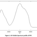 |
Figure 1: UV-Visible Spectrum profile of PFP. |
FT-IR Analysis of PFP
The spectrum of FT-IR can provide evidence of the extracts chemical composition. Results from the FT-IR spectrum, the functional groups such as polyphenolic compounds present in the PFP were distinguished from each other based on their peak ratio. Results obtained from the spectrum having 7 major peaks (Figure 2). The typical characteristics of phenolic compounds shown in the spectra are as follows: Peaks observed at 1605 cm⁻¹, 1515 cm⁻¹ and 1457 cm⁻¹ correspond to aromatic C=C bond vibrations, confirming the presence of aromatic rings. The peaks observed at 1375 cm⁻¹ corresponds to O-H bending vibration of the phenolic hydroxyl group and the peak observed at 968 cm⁻¹ suggests C-H bending of alkene in the trans conformation. Additionally, the sharp peaks at 2923 cm⁻¹ and 2853 cm⁻¹ corresponds to C-H stretching vibrations, which is the characteristic property of methoxy compounds. The presence of these methoxy groups indicates a diverse composition within the polyphenolic extract, potentially enhancing its functional properties.
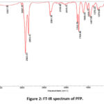 |
Figure 2: FT-IR spectrum of PFP. |
HPLC Analysis of PFP
In the present study, the HPLC analysis of PFP were carried out. The standard resveratrol showed 12.18 min retention time whereas the resveratrol peak appeared at the same retention time i.e. 12,13 min in the PFP sample. Additionally, the 42 % of resveratrol content was determined in PFP sample as per HPLC analysis. The results were shown in Figure 3.
 |
Figure 3: Representative chromatograms obtained from HPLC analysis. |
Anti-oxidant Activity of PFP by DPPH, FRAP, ABTS
The in vitro antioxidant activity of PFP was assessed in this study by evaluating its ability to scavenge free radicals, through the DPPH and ABTS assays, respectively, and its ability to reduce the iron(III) ion (Fe³⁺) in the FRAP assay. In the DPPH assay, PFP exhibited a higher scavenging capacity and enhanced antioxidant activity. Results indicated that the DPPH• scavenging activity of PFP increased significantly in a concentration dependent manner (Figure 4 (a)). Similarly, the effectiveness of PFP in scavenging ABTS+• is illustrated in Figure 4 (b). From the results, it clearly shown that PFP can have the ability to reduce the radical cation formation in a concentration dependent manner. PFP at higher concentration 100 µg/mL exhibited 83.53 % of inhibition. Similarly, in ABTS assay showed maximum radical scavenging activity at highest concentration (83.53 %). Furthermore, the FRAP assay is regarded as a reliable method for testing the antioxidant power of therapeutic compounds. Also, the FRAP activity of PFP was analysed based on its ability to reduce the Fe³⁺–TPTZ reagent. As shown in Figure 4 (c), PFP demonstrated the strongest reducing power, with FRAP values of 98 μM Fe(II)/g. The high FRAP value of PFP is likely due to the significant amount of polyphenol compounds present, as these compounds are known for their strong FRAP antioxidant activity. Hence, PFP has the capacity to reduce free radical formation, indicating its antioxidant activity and potential to mitigate oxidative stress.
Scavenging Activity of PFP on NO Production
In the present study, nitrite radical scavenging activity was conducted on PFP at concentrations ranging from 6.25 to 100 µg/mL and the percentage of free radical scavenging was plotted against the concentration of PFP was depicted in Figure 4 (d). Our findings revealed that the PFP inhibited the increased nitrite levels and thereby it scavenging the free radical production in a concentration dependent manner. At the higher concentration (100 µg/mL) PFP had its maximum inhibition (83.57 %). These results suggest that PFP is a strong radical scavenger.
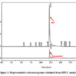 |
Figure 4: In vitro antioxidant enzymes (a) DPPH, (b) ABTS, (c) FRAP, (d) NO. Values are expressed as average of triplicate experiment and are represented as mean ± SD |
Anti-bacterial Analysis
In this study, PFP was evaluated against two Gram-positive (S. aureus, and MRSA) and four Gram-negative (E. coli, K. pneumoniae, P. mirabilis, P. auerogenisa) bacteria for determine the antibacterial effect. Results revealed that PFP had good anti-bacterial activity against both Gram-negative and Gram-positive and bacteria at different concentrations. Both S. aureus and MRSA showed higher zone of inhibition than standard drug Penicillin and Vancomycin. The maximum inhibition was obtained at the dose of 80 mg/mL and PFP showed higher inhibition as a concentration dependent increases. Moreover, PFP showed higher antibacterial activity against gram positive than against Gram negative bacteria. The results were shown in Table 2 and Figure 5.
Table 2: Anti-bacterial effect of PFP and its zone of inhibition.
|
Bacterial Strains |
Diameter of Inhibition Zone (mm) | |||
|
Concentration of PFP (µg/mL) |
||||
|
Gram Positive |
10 µg/mL | 20 µg/mL | 40 µg/mL | 80 µg/mL |
| S. aureus | 18.83 ±0.62 | 21.33±0.23 | 23.26±0.37 |
29.2±0.58 |
|
MRSA |
12.3±0.21 | 14.33±0.47 | 18±0.04 |
20.16±0.23 |
|
Gram Negative |
||||
|
E.coli |
10.5±0.40 | 13.33±0.47 | 14.36±0.44 | 16.8±0.86 |
| P. aeruginosa | 8.93±0.04 | 10.36±0.44 | 13.11±0.16 |
17.25±0.20 |
|
K. pneumonia |
11.06±0.09 | 14.16±0.23 | 15.06±0.09 | 18.4±0.43 |
| P. Mirabilis | 12±0.14 | 15.5±0.40 | 18.33±0.23 |
18.83±0.62 |
Values are expressed as average of triplicate experiment and are represented as mean ± SD
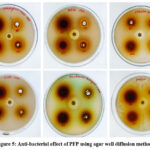 |
Figure 5: Anti-bacterial effect of PFP using agar well diffusion method. |
Discussion
Diverse phytochemicals that have evolved from plants and fruits are thought to be highly valuable as multifunctional substitutes for synthetic antioxidants in the reduction of oxidation in intricate food systems. These phytochemicals have a high level of natural antioxidant potential. Plant extracts have been subjected to phytochemical investigations, which have revealed the presence of components with physiological and medicinal properties.19 The antioxidant qualities of phenolic compounds found in different sections of different medicinal plants have been documented in numerous research. Antioxidants’ interaction with reactive free radicals is one of their primary roles. The main antioxidants that scavenge free radicals and reactive oxygen species are phenolic chemicals, such as stibilines, flavonoids and terpenoids. According to Larrauri,7 peanut skins are excellent precursors for the synthesis of polyphenolic compounds with strong antioxidant and chelating properties. Reducing expenses is a key component of converting waste into wealth, and this includes the peanut skin extracts, which are primarily flavonoids and polyphenols. The goal of this work was to extract resveratrol from the polyphenolics of peanut skin.
From UV-Vis spectrum profile of PFP demonstrated that the presence of polyphenols compounds due to peak shown at 300 nm with the maximum absorption of 1.401. The HPLC method was developed in this study to accurately estimate the amount of resveratrol in polyphenolic from peanut skin. A binary solvent system (acetonitrile in water and formic acid in water) was employed to achieve a sharp peak with negligible tailing and shorter retention time. Results demonstrated that resveratrol was detected at 306 nm with retention time 12.13 min in the PFP sample, similar to the standard resveratrol injection. Peaks observed at 1605 cm⁻¹, 1515 cm⁻¹ and 1457 cm⁻¹ correspond to aromatic C=C bond vibrations, confirming the presence of aromatic rings.20 The peaks observed at 1375 cm⁻¹ corresponds to O-H bending vibration of the phenolic hydroxyl group and the peak observed at 968 cm⁻¹ suggests C-H bending of alkene in the trans conformation, a distinctive feature of resveratrol.21 Additionally, the sharp peaks at 2923 cm⁻¹ and 2853 cm⁻¹ corresponds to C-H stretching vibrations, which is the characteristic property of methoxy compounds.22 The presence of these methoxy groups indicates a diverse composition within the polyphenolic extract, potentially enhancing its functional properties.
In the present study, the PFP had the ability to decolourize and scavenging the formation of free radicals. In vitro scavenging assays such as DPPH, ABTS, NO and FRAP were used to assessed the antioxidant capacity of PFP. The results obtained from this study revealed that TPC of PFP was 75 mg/GAE/g, suggesting that PPF has the ability to scavenge and may suppress the generation of reactive oxygen due to its unique properties.
The DPPH radical scavenging test is most common antioxidant assay that is dependent on radical scavenging activity, hydrogen donation, and/or substrate polarity.23 This approach is often considered indicative of plant extracts’ potential to scavenge free radicals, even though the DPPH radical has lesser significance to biological systems. It refers to hydrogen atom or electron donation ability, irrespective of any enzyme activity. Since DPPH radical is more persistent than hydroxyl or superoxide radicals, it is useful to employ this assay to assess the efficacy of antioxidants. Typically, a hydrogen atom transfer process is used in the DPPH test. Antioxidant molecules react with the radical to scavenge it by donating hydrogen, which is the mechanism by which antioxidants reduce the absorbance of DPPH. This appears as a discoloration that shifts from purple to yellow. The findings showed that PFP have 83.53 % radical inhibition ability. According to our findings, PFP antioxidant activity demonstrated a higher capacity for DPPH radical scavenging, suggesting a direct correlation between TPC and antioxidant activity. Numerous investigations have assessed the connection between plant products’ TPC and antioxidant activity. Antioxidants and radical scavengers are substances that have the ability to perform DPPH scavenging. Greater phenol content overall results in enhanced DPPH scavenging efficacy.
An additional method for assessing a sample’s capacity to scavenge radicals is the ABTS assay. Reactions with ABTS radicals often take place in less than a millisecond, and these radicals are more reactive than DPPH radicals. Both hydrogen-donating and chain-breaking antioxidants can be measured using ABTS radical cation decolorization, which can be used in both the organic and aqueous phases. Antioxidants in the extract have the ability to eliminate ABTS+ free radicals produced by a reaction with potassium persulfate. This is drastically at odds with the discolouration of a certain blue index and its ability to scavenge ABTS was expressed as an inhibition capability. Similar to the DPPH activity, the ABTS radical scavenging activity tended to rise in a concentration dependent manner. Furthermore, the extract may still retain the phenolic component profile linked to antioxidant action even when the polyphenol and ABTS levels were low. In this study, PFP showed 83.53% radical scavenging activity in a concentration dependent manner.
Another method utilized for the scavenging activity of PFP was FRAP analysis. This assay is based on the ability of an antioxidant to reduce the ferric tripyridyltriazine complex (Fe3 +-TPTZ) to the ferrous form intense blue Fe2 +-TPTZ with an absorption at 593 nm. Based on the decrease in absorbance, which is directly proportional to the antioxidant content present in the sample.24 In this study, the antioxidant activity of 98.53 µM/Fe (II)/mL was increased with increases in the PFP concentration. Additionally, the PFP capacity to scavenge the generation of nitric oxide (NO) was used to further analyse its radical scavenging potential. NO is crucial to the inflammatory process because it stimulates the oxidative damage, which leads to the generation of free radicals.25 According to our findings, PFP has a stronger NO scavenging effect at higher concentrations.
PFP had strong antioxidant activity because of its phenolic and other bioactive components, according to in vitro antioxidant tests. Consequently, this study was to ascertain PFP antibacterial activity by assessing the sensitivity of particular bacterial strains to PFP using the agar well diffusion method. When comparing PFP to the common antibiotics penicillin and vancomycin, notable growth inhibition of gram positive pathogens like S. aureus and MRSA was observed. Additionally, PFP exhibited comparatively more antibacterial effect against gram-positive organisms than gram-negative organisms. Altogether, this study highlighted that PFP has good anti-oxidant activity, which might result from elevated polyphenols concentrations. Consequently, it also exerts a significant anti-bacterial effect.
Conclusion
Overall results indicate that polyphenol isolated from peanut skin possess good anti-oxidant and anti-bacterial effects. Peanut skin extract contain resveratrol enriched polyphenol content with higher antioxidant activity, which is associated with increment in the free radical scavenging activity.
Acknowledgement
The author is profoundly grateful to Mr. Muralidhar Nayak, Research Scientist in Mass Spectrometry Facility at Indian Institute of Science (IISc.), Bangalore, India for the technical guidance.
Funding sources
The author(s) received no financial support for the research, authorship, and/or publication of this article
Conflict of Interest
The author(s) do not have any conflict of interest.
Data Availability Statement
This statement does not apply to this article.
Ethics Statement
This study did not involve human participants, animal subjects, or any material that requires ethical approval.
Informed Consent Statement
This study did not involve human participants, and therefore, informed consent was not required.
Authors’ Contribution
R. M. (Ratheesh Mohanan): Conceptualization, Methodology, Supervision, Writing- Review & Editing.
S. P. J. (Svenia Periyappurath Jose): Methodology, Data collection and Writing –Original Draft.
S. S (Sheethal Sreevallabhan): Data analysis, Visualization.
S. R. (Sony Rajan): Technical and material support.
References
- Sorita G.D., de Oliveira A., Moreira T.F.M., Leimann F.V., Ferreira S.R.S. Green-based processes applied for valorization of peanut by-product: In vitro evaluation of antioxidant and enzymatic inhibition capacities. Supercrit. Fluids. 2022; 186: 105602. DOI:10.1016/j.supflu.2022.105602.
CrossRef - Sorita G.D., Leimann F.V., Ferreira S.R.S. Biorefnery approach: Is it an upgrade opportunity for peanut by-products? Trends Food Sci. 2020; 105: 56–69. DOI: 1016/j.tifs.2020.08.011.
CrossRef - Sarnoski P.J., Boyer R.R., O’Keefe S.F. Application of proanthocyanidins from peanut skins as a natural yeast inhibitory agent. Food Sci. 2012; 77:242–24. DOI: 10.1111/j.1750-3841.2012.02652.x.
CrossRef - Win M.M., Abdul-Hamid A., Baharin B.S., Anwar F., Sabu M.C., Pak-Dek M.S. Phenolic compounds and antioxidant activity of peanuts skin, hull, raw kernel and roasted kernel flour. J. Bot. 2011; 43; 1635-1642.
- Xu M., Lv C., Wang H., Lu Q., Ye M., Zhu X., Liu R. Peanut skin extract ameliorates high-fat diet-induced atherosclerosis by regulating lipid metabolism, inflammation reaction and gut microbiota in ApoE−/− mice. Food Res Int. 2022; 154:111014. DOI: 1016/j.foodres.2022.111014.
CrossRef - Bazylko A., Parzonko A., Je W., Osinska E., Kiss A.K. Inhibition of ROS production, photoprotection, and total phenolic: flavonoids and ascorbic acid content of fresh herb juice and extracts from the leaves and flowers of Tropaeolum majus. Crops Prod. 2014; 55: 19–24.DOI10.1016/j.indcrop.2014.01.056.
CrossRef - Larrauri M., Zunino M.P., Zygadlo J.A., Grosso N.R., Nepote V. Chemical characterization and antioxidant properties of fractions separated from extract of peanut skin derived from different industrial processes. Ind Crops Prod. 2016; 94: 964-971. DOI:10.1016/j.indcrop.2016.09.066
CrossRef - Constanza K.E., White B.L., Davis J.P., Anders T.H., Dean L.L. Value-added processing of peanut skins: antioxidant capacity, total phenolics, and procyanidin content of spray-dried extracts. Agric. Food Chem. 2012; 60: 10776-10783. DOI: 10.1021/jf3035258.
CrossRef - Liu G., Shi A., Wang N., Li M., He X., Yin C., Tu Q., Shen X., Tao Y., Wang Q., Yin H. Polyphenolic proanthocyanidin-B2 suppresses proliferation of liver cancer cells and hepatocellular carcinogenesis through directly binding and inhibiting AKT activity. Redox Biol. 2020; 37: 101701. DOI: 1016/j.redox.2020.101701.
CrossRef - Rauf A., Imran M., Abu-Izneid T., Iahtisham-Ul-Haq., Patel S., Pan X., Naz S., Sanches S.A., Saeed F., Rasul S.H.A. Proanthocyanidins: A comprehensive review. Pharmacother. 2019; 116. DOI: 10.1016/j.biopha.2019.108999.
CrossRef - Unusan N. Proanthocyanidins in grape seeds: An updated review of their health benefits and potential uses in the food industry. Funct. Foods. 2020; 67: 103861. DOI: 10.1016/j.jff.2020.103861.
CrossRef - De Camargo A.C., Regitano-d’Arce M.A.B., Rasera G.B., Canniatti-Brazaca S.G., do Prado Silva L., Alvarenga V.O., Sant’Ana A.S., Shahidi F. Phenolic acids and flavonoids of peanut by-products: Antioxidant capacity and antimicrobial effects. Food Chem. 2017; 237: 538-544. DOI: 1016/j.foodchem.2017.05.046.
CrossRef - Harborne J.B. Phytochemical Methods. Chapman and Hall Ltd., London, 1973; 49-188.
- Velioglu Y.S., Mazza G., Gao L., Oomah B.D. Antioxidant activity and total phenolics in selected fruits, vegetables, and grain products. Agric. Food Chem.1998; 46: 4113-4117. DOI: http://dx.doi.org/10.1021/jf9801973.
CrossRef - Huang D., Ou B., Prior R.L. The chemistry behind antioxidant capacity assays. JAgric Food Chem. 2005; 53: 1841–56. DOI: https://doi.org/10.1021/jf030723c.
CrossRef - Re R., Pellegrini N., Proteggente A., Yang M., Rice-Evans C. Antioxidant activity applying an improved ABTS radical cation decolorization assay. Free Radic Biol 1999; 26 1231–7. DOI: 10.1016/s0891-5849(98)00315-3.
CrossRef - Benzie I.F, Strain J.J. The ferric reducing ability of plasma (FRAP) as measurement of “antioxidant power” The FRAP assay. Anal Biochem. 1996; (15) 239:70–6. DOI: 1006/abio.1996.0292.
CrossRef - Kamble S.C., Humbare R.B., Sarkar J., Kulkarni A.A. Assessment of phytochemicals and antioxidant properties of root extracts of Rubia cordifolia Sciforum. Biol Life Sci Forum 2021; 4:100. DOI: https://doi.org/10.3390/IECPS2020-08625.
CrossRef - Fresco P., Borges F., Diniz C., Marques M.P. New insights on the an-ticancer properties of dietary polyphenols. Med Res Rev 2006; 26: 747-66. DOI:1002/med.20060.
CrossRef - Li D., Wei Z., Li X. Development, characterization and resveratrol delivery of hollow gliadin nanoparticles: advantages over solid gliadin nanoparticles. Foods 2023; 12: 2436. DOI: https://doi.org/10.3390/foods12132436.
CrossRef - Porto I.C.C.M., Nascimento T.G., Oliveira J.M.S., Freitas P.H., Haimeur A., França R. Use of polyphenols as a strategy to prevent bond degradation in the dentin–resin interface. Eur J Oral Sci. 2018; 126:146-158. DOI: 1111/eos.12403.
CrossRef - Sim C., Hamdan M.R., Ismail Z., Ahmad M. Assessment of herbal medicines by chemometrics-assisted interpretation of FTIR spectra. J Analytica Chimica Acta. 2004;1: 14.
- Milardović S., Iveković D., Grabarić B.S. A novel amperometric method for antioxidant activity determination using DPPH free radical. 2006; 68:175–80. DOI:10.1016/j.bioelechem.2005.06.005.
CrossRef - Liu L., Sun Y., Laura T., Liang X., Ye H., Zeng X. Determination of polyphenolic content and antioxidant activity of kudingcha made from Ilex kudingcha CJ Tseng. Food Chem. 2009; 112:35-41.DOI:1016/j.foodchem.2008.05.038.
CrossRef - Mfotie Njoya E, Munvera AM, Mkounga P, Nkengfack AE, McGaw LJ. Phytochemical analysis with free radical scavenging, nitric oxide inhibition and antiproliferative activity of Sarcocephalus pobeguiniiBMC Complement Altern Med. 2017; 17:199. DOI: 10.1186/s12906-017-1712-5.
CrossRef

This work is licensed under a Creative Commons Attribution 4.0 International License.